Overexpression of LMO4 induces mammary hyperplasia, promotes cell invasion, and is a predictor of poor outcome in breast cancer
- PMID: 15897450
- PMCID: PMC1140463
- DOI: 10.1073/pnas.0502990102
Overexpression of LMO4 induces mammary hyperplasia, promotes cell invasion, and is a predictor of poor outcome in breast cancer
Abstract
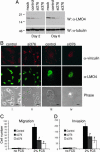
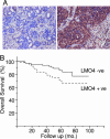
The zinc finger protein LMO4 is overexpressed in a high proportion of breast carcinomas. Here, we report that overexpression of a mouse mammary tumor virus (MMTV)-Lmo4 transgene in the mouse mammary gland elicits hyperplasia and mammary intraepithelial neoplasia or adenosquamous carcinoma in two transgenic strains with a tumor latency of 13-18 months. To investigate cellular processes controlled by LMO4 and those that may be deregulated during oncogenesis, we used RNA interference. Down-regulation of LMO4 expression reduced proliferation of human breast cancer cells and increased differentiation of mouse mammary epithelial cells. Furthermore, small-interfering-RNA-transfected breast cancer cells (MDA-MB-231) had a reduced capacity to migrate and invade an extracellular matrix. Conversely, overexpression of LMO4 in noninvasive, immortalized human MCF10A cells promoted cell motility and invasion. Significantly, in a cohort of 159 primary breast cancers, high nuclear levels of LMO4 were an independent predictor of death from breast cancer. Together, these findings suggest that deregulation of LMO4 in breast epithelium contributes directly to breast neoplasia by altering the rate of cellular proliferation and promoting cell invasion.
Figures


Similar articles
-
The LIM-only factor LMO4 regulates expression of the BMP7 gene through an HDAC2-dependent mechanism, and controls cell proliferation and apoptosis of mammary epithelial cells.Oncogene. 2007 Sep 27;26(44):6431-41. doi: 10.1038/sj.onc.1210465. Epub 2007 Apr 23. Oncogene. 2007. PMID: 17452977
-
The LIM domain gene LMO4 inhibits differentiation of mammary epithelial cells in vitro and is overexpressed in breast cancer.Proc Natl Acad Sci U S A. 2001 Dec 4;98(25):14452-7. doi: 10.1073/pnas.251547698. Proc Natl Acad Sci U S A. 2001. PMID: 11734645 Free PMC article.
-
Loss of the LIM domain protein Lmo4 in the mammary gland during pregnancy impedes lobuloalveolar development.Oncogene. 2005 Jul 14;24(30):4820-8. doi: 10.1038/sj.onc.1208638. Oncogene. 2005. PMID: 15856027
-
[Involvement of LMO4 in tumorigenesis associated epithelial-mesenchymal transition].Zhejiang Da Xue Xue Bao Yi Xue Ban. 2011 Jan;40(1):107-11. doi: 10.3785/j.issn.1008-9292.2011.01.019. Zhejiang Da Xue Xue Bao Yi Xue Ban. 2011. PMID: 21319383 Review. Chinese.
-
Cell cycle genes in a mouse mammary hyperplasia model.J Mammary Gland Biol Neoplasia. 2004 Jan;9(1):81-93. doi: 10.1023/B:JOMG.0000023590.63974.f8. J Mammary Gland Biol Neoplasia. 2004. PMID: 15082920 Review.
Cited by
-
LMO4 mediates trastuzumab resistance in HER2 positive breast cancer cells.Am J Cancer Res. 2018 Apr 1;8(4):594-609. eCollection 2018. Am J Cancer Res. 2018. PMID: 29736306 Free PMC article.
-
Discovery of novel mechanosensitive genes in vivo using mouse carotid artery endothelium exposed to disturbed flow.Blood. 2010 Oct 14;116(15):e66-73. doi: 10.1182/blood-2010-04-278192. Epub 2010 Jun 15. Blood. 2010. PMID: 20551377 Free PMC article.
-
Requirement for Lmo4 in the vestibular morphogenesis of mouse inner ear.Dev Biol. 2010 Feb 1;338(1):38-49. doi: 10.1016/j.ydbio.2009.11.003. Epub 2009 Nov 10. Dev Biol. 2010. PMID: 19913004 Free PMC article.
-
LMO4 is an essential cofactor in the Snail2-mediated epithelial-to-mesenchymal transition of neuroblastoma and neural crest cells.J Neurosci. 2013 Feb 13;33(7):2773-83. doi: 10.1523/JNEUROSCI.4511-12.2013. J Neurosci. 2013. PMID: 23407937 Free PMC article.
-
Nimodipine Used with Vincristine: Protects Schwann Cells and Neuronal Cells from Vincristine-Induced Cell Death but Increases Tumor Cell Susceptibility.Int J Mol Sci. 2024 Sep 27;25(19):10389. doi: 10.3390/ijms251910389. Int J Mol Sci. 2024. PMID: 39408743 Free PMC article.
References
-
- Rabbitts, T. H. (1998) Genes Dev. 12, 2651–2657. - PubMed
Publication types
MeSH terms
Substances
LinkOut - more resources
Full Text Sources
Other Literature Sources
Medical
Molecular Biology Databases
Miscellaneous

