Comparative Study
. 2005 May 24;102(21):7505-10.
doi: 10.1073/pnas.0409482102. Epub 2005 May 16.

The crystal structures of human steroidogenic factor-1 and liver receptor homologue-1

Affiliations
Comparative Study

The crystal structures of human steroidogenic factor-1 and liver receptor homologue-1

Weiru Wang et al. Proc Natl Acad Sci U S A. .

Abstract

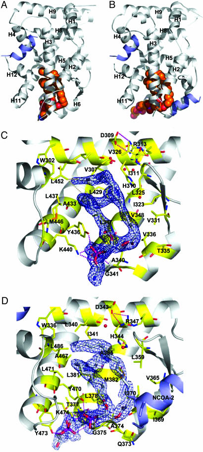
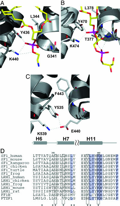
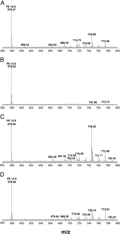
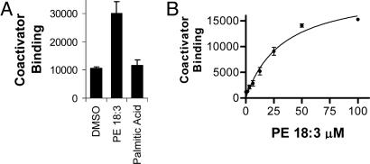
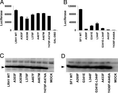
Steroidogenic factor-1 (SF-1) and liver receptor homologue-1 (LRH-1) belong to the fushi tarazu factor 1 subfamily of nuclear receptors. SF-1 is an essential factor for sex determination during development and regulates adrenal and gonadal steroidogenesis in the adult, whereas LRH-1 is a critical factor for development of endodermal tissues and regulates cholesterol and bile acid homeostasis. Regulatory ligands are unknown for SF-1 and LRH-1. A reported mouse LRH-1 structure revealed an empty pocket in a region commonly occupied by ligands in the structures of other nuclear receptors, and pocket-filling mutations did not alter the constitutive activity observed. Here we report the crystal structures of the putative ligand-binding domains of human SF-1 at 2.1-A resolution and human LRH-1 at 2.5-A resolution. Both structures bind a coactivator-derived peptide at the canonical activation-function surface, thus adopting the transcriptionally activating conformation. In human LRH-1, coactivator peptide binding also occurs to a second site. We discovered in both structures a phospholipid molecule bound in a pocket of the putative ligand-binding domain. MS analysis of the protein samples used for crystallization indicated that the two proteins associate with a range of phospholipids. Mutations of the pocket-lining residues reduced the transcriptional activities of SF-1 and LRH-1 in mammalian cell transfection assays without affecting their expression levels. These results suggest that human SF-1 and LRH-1 may be ligand-binding receptors, although it remains to be seen if phospholipids or possibly other molecules regulate SF-1 or LRH-1 under physiological conditions.


Figures

Fig. 1.
Fig. 1.
The hSF-1 and hLRH-1 LBD structures complexed with phospholipid and coactivator peptide. (A and B) The hSF-1 LBD (A) and the hLRH-1 LBD (B) (gray ribbon models) with phospholipid ligands (spherical model colored by atom type), and NCOA2 coactivator peptide (blue ribbon model). Note that two NCOA2 peptides bind to each LRH-1 molecule, one at the canonical activation function surface and the other at a site formed by H2, H3, and the β-sheet. (C) Residues of the hSF-1 ligand-binding pocket (stick models colored yellow and by atom type), showing salt bridge and hydrogen bonds (dotted lines) to the PE (stick models colored white and by atom type). The blue mesh indicates an unbiased 2FoFc map covering the ligand. H2 and H3 are truncated to show the pocket features. (D) Residues of the hLRH-1 ligand-binding pocket depicted as in C showing interactions with the phosphatidylglycerol-phosphoglycerol.
Fig. 2.
Fig. 2.
Comparison of hSF-1 and hLRH-1 structures with mLRH-1. (A and B) A phosphate group in hSF-1 (A) and hLRH-1 (B) interacts with the Lys and Tyr of the KYG triad. (C) E440 in the APO mLRH-1 mimics the phosphate group interactions. Only the residues of the phosphate-binding triad (sticks colored yellow and by atom type) and the polar portions of the phospholipids (sticks colored by atom type) are shown. (D) Alignment of representative NR5A sequences. The two sequence segments that contain the KYG motif (helix H6–H7 and H11) are shown. A more complete alignment appears in Fig. 8.
Fig. 3.
Fig. 3.
Mass spectral analysis of lipids bound to hSF-1 and hLRH-1 LBD proteins purified from E. coli: wild-type hSF-1 (A), hSF-1 Y436F-K440A (B), wild-type hLRH-1 (C), and hLRH-1 Y470F-K474A (D). The analyses were performed in negative mode. PE-12:0 (50 pmol) was mixed with 50 pmol of each LBD protein before extraction, giving the m/z = 578 standard peak.
Fig. 4.
Fig. 4.
PE dose-dependent increase in coactivator recruitment to hSF-1 in vitro. (A) PE-18:3 (50 μM 1,2-dilinolenoyl-sn-glycero-3-phosphoethanolamine) but not palmitic acid (50 μM) activates wild-type hSF-1 to bind NCOA1 by AlphaScreen. (B) Dose-dependent NCOA1 recruitment to hSF-1 by PE-18:3. Error bars indicate the standard deviations. The graphs shown are representative of three experiments.
Fig. 5.
Fig. 5.
Effects of pocket mutations on hLRH-1 and hSF-1 functions in HEK293T cells. (A) hLRH-1 LBD activity tested as GAL-DBD fusions acting at a GAL4-responsive LUC reporter gene. The mutations tested include residues A303, L378, A467, Y470, and K474. (B) hSF-1 LBD activity tested as GAL-DBD fusions. The mutations tested include residues A269, G341, L344, A433, Y436, and K440. (C) Western blot analysis of cells after transfection with vectors encoding GAL4-DBD–hLRH-1 LBD fusion proteins using anti-GAL4-DBD antibody. (D) Western blot analysis of GAL4-DBD hSF-1 LBD fusion proteins. Error bars indicate the standard deviations. The graphs shown are representative of three experiments.

References

    1. Lavorgna, G., Ueda, H., Clos, J. & Wu, C. (1991) Science 252, 848–851. - PubMed
    1. Chai, C. & Chan, W. K. (2000) Mech. Dev. 91, 421–426. - PubMed
    1. Laudet, V. (1997) J. Mol. Endocrinol. 19, 207–226. - PubMed
    1. Ueda, H., Sun, G. C., Murata, T. & Hirose, S. (1992) Mol. Cell. Biol. 12, 5667–5672. - PMC - PubMed
    1. Parker, K. L. & Schimmer, B. P. (1997) Endocr. Rev. 18, 361–377. - PubMed

Publication types

MeSH terms

LinkOut - more resources