Comparative Study
. 2005 May 31;102(22):7988-93.
doi: 10.1073/pnas.0409465102. Epub 2005 May 16.

Human coronavirus NL63 employs the severe acute respiratory syndrome coronavirus receptor for cellular entry

Affiliations
Comparative Study

Human coronavirus NL63 employs the severe acute respiratory syndrome coronavirus receptor for cellular entry

Heike Hofmann et al. Proc Natl Acad Sci U S A. .

Abstract

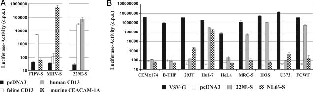
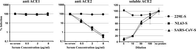
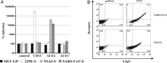
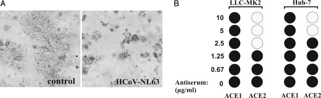
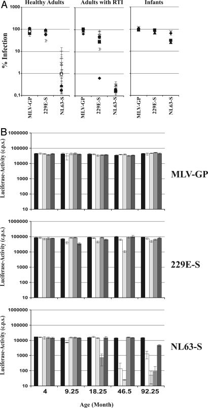
Coronavirus (CoV) infection of humans is usually not associated with severe disease. However, discovery of the severe acute respiratory syndrome (SARS) CoV revealed that highly pathogenic human CoVs (HCoVs) can evolve. The identification and characterization of new HCoVs is, therefore, an important task. Recently, a HCoV termed NL63 was discovered in patients with respiratory tract illness. Here, cell tropism and receptor usage of HCoV-NL63 were analyzed. The NL63 spike (S) protein mediated infection of different target cells compared with the closely related 229E-S protein but facilitated entry into cells known to be permissive to SARS-CoV-S-driven infection. An analysis of receptor engagement revealed that NL63-S binds angiotensin-converting enzyme (ACE) 2, the receptor for SARS-CoV, and HCoV-NL63 uses ACE2 as a receptor for infection of target cells. Potent neutralizing activity directed against NL63- but not 229E-S protein was detected in virtually all sera from patients 8 years of age or older, suggesting that HCoV-NL63 infection of humans is common and usually acquired during childhood. Here, we show that SARS-CoV shares its receptor ACE2 with HCoV-NL63. Because the two viruses differ dramatically in their ability to induce disease, analysis of HCoV-NL63 might unravel pathogenicity factors in SARS-CoV. The frequent HCoV-NL63 infection of humans suggests that highly pathogenic variants have ample opportunity to evolve, underlining the need for vaccines against HCoVs.


Figures

Fig. 1.
Fig. 1.
Cellular tropism of NL63-S- and 229E-S-bearing pseudotypes. (A) 293T cells were transfected with CoV receptors or pcDNA3 and infected with the indicated viral pseudotypes, and luciferase activities in the cell lysates were determined. c.p.s., counts per sec. A representative experiment is shown. Comparable results were obtained in an independent experiment. Error bars indicate SD. (B) Cell lines were infected with the indicated p24-normalized viral pseudotypes, and luciferase activities in the cell lysates were determined. The results were confirmed in three independent experiments. Error bars indicate SD.
Fig. 2.
Fig. 2.
Inhibition of NL63-S-driven infection by ACE2-specific antibodies and soluble ACE2. Huh-7 cells were preincubated with ACE1- (Left) or ACE2- (Center) specific polyclonal antibodies, or the pseudotyped virions were preincubated with the ACE2 ectodomain (Right). Subsequently, the Huh-7 cells were infected with the indicated pseudovirions, and luciferase activities in the cell lysates were quantified. The results are shown as the percent of infection in the absence of inhibitor and were confirmed in two independent experiments. Error bars indicate SD.
Fig. 3.
Fig. 3.
Expression of ACE2 potentiates NL63-S-driven infection, and soluble NL63-S protein binds to ACE2-positive cells. (A) 293T cells expressing CD13, ACE2, ACE1, or pcDNA3 were infected with the indicated pseudotypes, and luciferase activities in the cell lysates were determined. The results are shown as the percent of infection of pcDNA3-transfected cells. Similar results were obtained in three independent experiments. Error bars indicate SD. (B) ACE2 or pcDNA3 were transiently expressed on 293T cells, the cells were incubated with the S1 subunit of NL63-S or SARS-CoV-S fused to the Fc portion of human immunoglobulin, and receptor expression and S-Fc-fusion-protein binding were analyzed by FACS. Two independent experiments yielded similar results. NL63- and SARS-CoV-S binding was assessed with the same batch of transfected cells. Differences in ACE2 signal might be due to the masking of different ACE2 epitopes by the two S proteins, resulting in differential recognition of ACE2 by the polyclonal serum.
Fig. 4.
Fig. 4.
Inhibition of HCoV-NL63 replication by ACE2-specific antibodies. (A) Huh-7 cells were infected with HCoV-NL63 or mock infected, and CPE development was assessed 5 d after infection. Comparable results were obtained in several independent experiments. (B) LLC-MK2 and Huh-7 cells were preincubated with the indicated concentrations of ACE1- or ACE2-specific polyclonal antibodies and infected with HCoV-NL63, and the development of CPE was assessed. •, CPE development; ○, absence of CPE. Similar results were obtained in an independent experiment.
Fig. 5.
Fig. 5.
Neutralization of NL63-S-driven infection by human sera. (A) The indicated pseudovirions were incubated with 50-fold-diluted sera from healthy adults, adults with RTI, or infants and added onto Huh-7 cells, and luciferase activities in the cell lysates were determined. The results were confirmed in an independent experiment. Error bars indicate SD. (B) The indicated pseudovirions were incubated with 50-fold-diluted sera from a total of 25 infants of defined age groups and used for infection of Huh-7 cells as described for A. Sera from four individuals were analyzed per age group. Within age groups, bars indicate results obtained with serum from single individuals. The black bars indicate infection in the absence of patient serum. An independent experiment yielded similar results. Error bars indicate SD.

Similar articles

Cited by

  • Polyunsaturated ω-3 fatty acids inhibit ACE2-controlled SARS-CoV-2 binding and cellular entry.
    Goc A, Niedzwiecki A, Rath M. Goc A, et al. Sci Rep. 2021 Mar 4;11(1):5207. doi: 10.1038/s41598-021-84850-1. Sci Rep. 2021. PMID: 33664446 Free PMC article.
  • Cross-Sectional Evaluation of Humoral Responses against SARS-CoV-2 Spike.
    Prévost J, Gasser R, Beaudoin-Bussières G, Richard J, Duerr R, Laumaea A, Anand SP, Goyette G, Benlarbi M, Ding S, Medjahed H, Lewin A, Perreault J, Tremblay T, Gendron-Lepage G, Gauthier N, Carrier M, Marcoux D, Piché A, Lavoie M, Benoit A, Loungnarath V, Brochu G, Haddad E, Stacey HD, Miller MS, Desforges M, Talbot PJ, Maule GTG, Côté M, Therrien C, Serhir B, Bazin R, Roger M, Finzi A. Prévost J, et al. Cell Rep Med. 2020 Oct 20;1(7):100126. doi: 10.1016/j.xcrm.2020.100126. Epub 2020 Sep 30. Cell Rep Med. 2020. PMID: 33015650 Free PMC article.
  • Structural Analysis of the OC43 Coronavirus 2'-O-RNA Methyltransferase.
    Dostalik P, Krafcikova P, Silhan J, Kozic J, Chalupska D, Chalupsky K, Boura E. Dostalik P, et al. J Virol. 2021 Jul 12;95(15):e0046321. doi: 10.1128/JVI.00463-21. Epub 2021 Jul 12. J Virol. 2021. PMID: 34011548 Free PMC article.
  • Potential use of renin-angiotensin-aldosterone system inhibitors to reduce COVID-19 severity.
    Gonçalves J, Santos CD, Fresco P, Fernandez-Llimos F. Gonçalves J, et al. Rev Port Cardiol. 2023 Apr;42(4):373-383. doi: 10.1016/j.repc.2022.02.014. Epub 2023 Mar 7. Rev Port Cardiol. 2023. PMID: 36893838 Free PMC article. Review.
  • The Omic Insights on Unfolding Saga of COVID-19.
    Kaur A, Chopra M, Bhushan M, Gupta S, Kumari P H, Sivagurunathan N, Shukla N, Rajagopal S, Bhalothia P, Sharma P, Naravula J, Suravajhala R, Gupta A, Abbasi BA, Goswami P, Singh H, Narang R, Polavarapu R, Medicherla KM, Valadi J, Kumar S A, Chaubey G, Singh KK, Bandapalli OR, Kavi Kishor PB, Suravajhala P. Kaur A, et al. Front Immunol. 2021 Oct 20;12:724914. doi: 10.3389/fimmu.2021.724914. eCollection 2021. Front Immunol. 2021. PMID: 34745097 Free PMC article. Review.

References

    1. Holmes, K. V. (2001) in Fields Virology, eds. Fields, B. N., Knipe, D. M., Howley, P. M., Griffin, D. E., Lamb, R. A., Martin, M. A., Roizman, B. & Straus, S. E. (Lippincott, Williams & Wilkins, Philadelphia), pp. 1187–1203.
    1. Stadler, K., Masignani, V., Eickmann, M., Becker, S., Abrignani, S., Klenk, H. D. & Rappuoli, R. (2003) Nat. Rev. Microbiol. 1, 209–218. - PMC - PubMed
    1. Peiris, J. S., Yuen, K. Y., Osterhaus, A. D. & Stohr, K. (2003) N. Engl. J. Med. 349, 2431–2441. - PubMed
    1. Gallagher, T. M. & Buchmeier, M. J. (2001) Virology 279, 371–374. - PMC - PubMed
    1. Kuo, L., Godeke, G. J., Raamsman, M. J., Masters, P. S. & Rottier, P. J. (2000) J. Virol. 74, 1393–1406. - PMC - PubMed

Publication types

MeSH terms

LinkOut - more resources