. 2005 Jul 7;24(29):4728-35.
doi: 10.1038/sj.onc.1208674.

Interaction and colocalization of Rad9/Rad1/Hus1 checkpoint complex with replication protein A in human cells

Affiliations

Interaction and colocalization of Rad9/Rad1/Hus1 checkpoint complex with replication protein A in human cells

Xiaoming Wu et al. Oncogene. .

Abstract

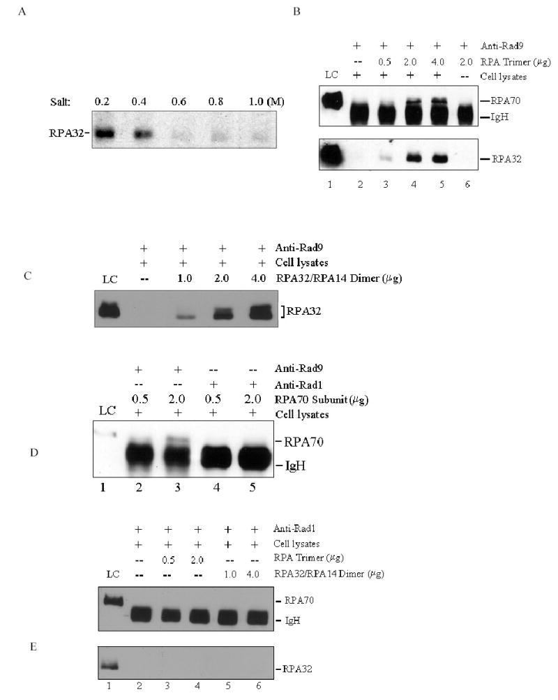
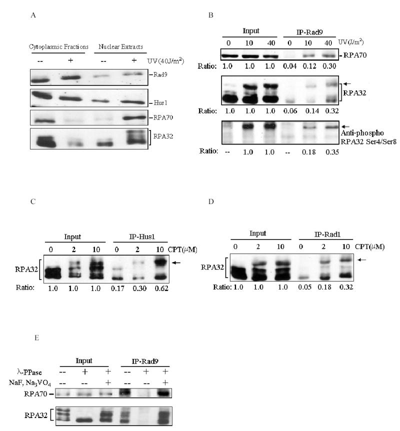
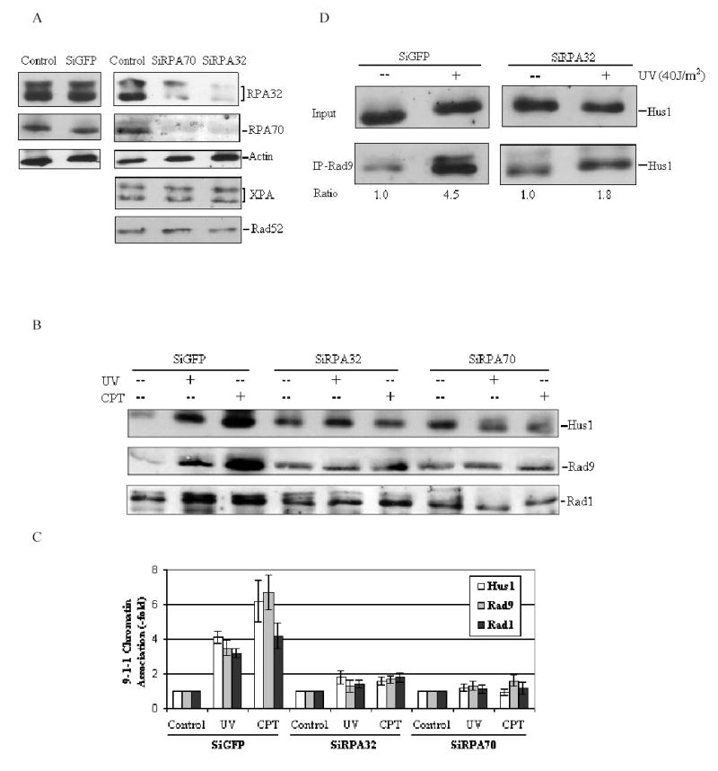
Replication protein A (RPA) is a eukaryotic single-stranded DNA-binding protein consisting of three subunits of 70-, 32-, and 14-kDa (RPA70, RPA32, RPA14, respectively). It is a protein essential for most cellular DNA metabolic pathways. Checkpoint proteins Rad9, Rad1, and Hus1 form a clamp-like complex which plays a central role in the DNA damage-induced checkpoint response. In this report, we presented the evidence that Rad9-Rad1-Hus1 (9-1-1) complex directly interacted with RPA in human cells, and this interaction was mediated by the binding of Rad9 protein to both RPA70 and RPA32 subunits. In addition, the cellular interaction of 9-1-1 with RPA or hyperphosphorylated RPA was stimulated by UV irradiation or camptothecin treatment in a dose-dependent manner. Such treatments also resulted in the colocalization of the nuclear foci formed with the two complexes. Consistently, knockdown of the RPA expression in cells by the small interference RNA (siRNA) blocked the DNA damage-dependent chromatin association of 9-1-1, and also inhibited the 9-1-1 complex formation. Taken together, our results suggest that 9-1-1 and RPA complexes collaboratively function in DNA damage responses, and that the RPA may serve as a regulator for the activity of 9-1-1 complex in the cellular checkpoint network.


Figures

Figure 1
Figure 1
Interaction of RPA with Rad9-Rad1-Hus1 complex. (A): Total cell lysates prepared from exponentially growing HeLa cells were used for co-immunoprecipitation (IP) assays with anti-Rad9 (lane 2), anti-Rad1 (lane 3) and anti-Hus1 (lane 4) or normal rabbit IgG (lane 5) as described in Materials and methods. Proteins from the immunoprecipitates were detected by Western blotting using anti-RPA70 antibody (upper panel) or anti-RPA32 antibody (lower panel). As control, 10% of the total volumes of the whole cellular lysates used for the co-IP reactions were also included (Input, lane 1). IgH is the IgG heavy chain. (B): Total cell lysates were subjected to the co-IP assays with anti-RPA70 antibody and the immunoprecipitated proteins were detected using anti-Rad1 (upper panel) or anti-Hus1 antibody (lower panel). (C): Cell lysates were either treated with 100 μg/mL DNase I (Invitrogen) for 20 min at 37 °C (lane 2) or 50 μg/mL ethidium bromide (EtBr) on ice for 30 min (lane 3) or mock-treated (lane 1). Cell lysates were then subjected to co-IP assays with anti-Rad9 antibody and detected by anti-RPA32 antibody (lower panel). 10% of treated cell lysates were also included as controls (upper panel). Data are representative of three independent experiments.
Figure 2
Figure 2
Interaction of Rad9-Rad1-Hus1 complex with RPA is mediated by Rad9. (A): Total cellular lysates were incubated with anti-Rad9 antibodies for 10–14 h, followed by 1-h incubation with protein A/G-agarose beads. After centrifugation, the immunoprecipitates were washed three times with PBS containing 0.05% Nonidet P-40, and further incubated with increasing salt concentrations of buffer (15 mM Tris-Cl, pH 7.5, 0.2~1.0 M NaCl, 0.1% NP-40) for 30 min at 4 °C. After washing, the remaining bound proteins were detected by anti-RPA32 antibody. (B): The co-IP reactions were performed as in Fig. 2A, and the immunoprecipitates were then incubated with the buffer containing 0.6 M NaCl. After centrifugation and washing, purified RPA was added and further incubated in 500 μL RPA binding buffer (40 mM HEPES-KOH, pH 7.5, 75 mM KCl, 8 mM MgCl2, 1 mM DTT, 5% glycerol and 100 μg/mL BSA, 0.1% NP-40) for 4-6 h. The bound proteins were detected by Western blotting with anti-RPA70 and anti-RPA32 antibodies, respectively. (C): The experiments were conducted similarly as described in Fig. 2B, except that RPA32/RPA14 subunit was finally added. (D): The experiments were conducted similarly as described in Fig. 2B, except that anti-Rad9 antibody (lanes 2 and 3) and anti-Rad1 antibody (lanes 4 and 5) were used for co-IP reactions and RPA70 subunit was finally added. (E): The experiments were conducted similarly as described in Fig. 2B, but anti-Rad1 antibody was used for co-IP reactions, and either RPA trimer (lanes 3 and 4) or RPA32/RPA14 subunit (lanes 5 and 6) was finally added. 10% of purified protein used for final binding assays was loaded as control (LC: loading control). Data are representative of two independent experiments.
Figure 3
Figure 3
DNA damage stimulates Rad9 checkpoint complex interaction with RPA. (A): Cells were treated with indicated doses of UV irradiation followed by a 2-h recovery. Nuclear extracts were prepared and separated on SDS-PAGE gels, and then probed with anti-Rad9, anti-Rad1, anti-RPA70 and anti-RPA32 antibodies, respectively. (B): Cells were treated with indicated doses of UV and total cell lysates were used for co-IP assays with anti-Rad9 antibody. The immunoprecipitates were then analyzed with anti-RPA70 (upper panel), anti-RPA32 (middle panel) and anti-phospho-RPA32 Ser4/Ser8 (lower panel) antibodies, respectively. The arrows indicate the hyperphosphorylated RPA. The relative quantities of proteins immunoprecipitated by anti-Rad9 antibody were estimated by densitometry and further normalized based on input, which were designated as 1.0. (C) and (D): Cells were treated with indicated doses of CPT, and then the total cell lysates were used for co-IP assays with anti-Hus1(C) or anti-Rad1 (D) antibodies. The immunoprecipitates were detected with anti-RPA32 antibody. (E): Whole cellular lysates were prepared from 40 J/m2 UV-irradiated cells. Prior to co-IP assays, the lysates were either mock treated (−/−), or treated with 2000 units of λ-phosphatase (λ-PPase) (New England Biolabs) for 30 min at 30 °C in the absence (−/+) or presence (+/+) of 10 mM of NaF and 1 mM Na3VO4. Treated cell lysates were then subjected for co-IP reactions with anti-Rad9 antibody. The bound proteins were then detected by Western blotting using anti-RPA32 antibody. Data are representative of at least three independent experiments.
Figure 4
Figure 4
Co-localization of RPA with 9-1-1 complex. Cells were treated with 40 J/m2 UV irradiation or 10 μM CPT. After extraction of cytoplasmic proteins with PBS containing 0.5% NP-40, cells were fixed and incubated with primary and secondary antibodies, and visualized with fluorescent microscopy. (A): Cells were stained with anti-Rad9 antibody (green, subpanels B, F, and J) and anti-RPA32 antibody (red, subpanels C, G, and K). Subpanels D, H and L are the merged images of the anti-Rad9 and anti-RPA32 stained cells. Subpanels A, E and I are the DAPI (4′,6-diamidino-2-phenylindole)-stained nuclei. (B): Cells were stained with anti-Rad1 antibody (red, subpanels B, F, and J) and anti-phospho-RPA32 Ser4/Ser8 antibody (green, subpanels C, G, and K). Subpanels D, H and L are the merged images of the anti-Rad1 and anti-phospho-RPA32 Ser4/Ser8 stained cells.
Figure 5
Figure 5
Knockdown of RPA by siRNA affects 9-1-1 complex accumulation on chromatin following DNA damage. (A): HeLa cells were transfected with siRPA70, siRPA32 or siGFP as described in Materials and methods. Controls were treated with transfection reagents only. Total cell lysates were harvested 72 h after transfection, and probed with indicated antibodies, respectively. (B): HeLa cells were transfected with indicated siRNA, and then cells were treated with 40 J/m2 UV irradiation or 10μM CPT at 72h after transfection. The chromatin-bound fractions were isolated as described in materials and methods and immunoblotted with anti-Rad9, anti-Rad1 and anti-Hus1 antibodies, respectively. (C): The data obtained from panel (B) were quantified using Fuji Image Gauge 3.46, and normalized to the controls (as the value of 1) and are the mean ± SD of three independent experiments. (D): HeLa cells were transfected with siRPA32 or siGFP, and then treated with 40 J/m2 UV irradiation 72 h after transfection. The nuclear extracts were prepared and used for co-IP assays with anti-Rad9 antibody. The bound protein was detected with anti-Hus1 antibody. The quantity of Hus1 immunoprecipitated by anti-Rad9 antibody was normalized to input.

References

    1. Abraham RT. Genes Dev. 2001;15:2177–2196. - PubMed
    1. Bao S, Lu T, Wang X, Zheng H, Wang LE, Wei Q, Hittelman WN, Li L. Oncogene. 2004;23:5586–5593. - PubMed
    1. Barr SM, Leung CG, Chang EE, Cimprich KA. Curr Biol. 2003;13:1047–1051. - PubMed
    1. Bartek J, Lukas C, Lukas J. Nat Rev Mol Cell Biol. 2004;5:792–804. - PubMed
    1. Bermudez VP, Lindsey-Boltz LA, Cesare AJ, Maniwa Y, Griffith JD, Hurwitz J, Sancar A. Proc Natl Acad Sci USA. 2003;100:1633–1638. - PMC - PubMed

Publication types