Review
. 2005 Apr;2(2):333-47.
doi: 10.1602/neurorx.2.2.333.

Imaging in neurooncology

Affiliations
Review

Imaging in neurooncology

Andreas H Jacobs et al. NeuroRx. 2005 Apr.

Abstract

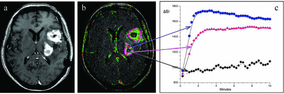
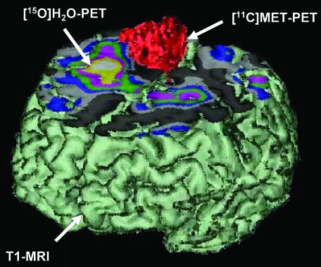
Imaging in patients with brain tumors aims toward the determination of the localization, extend, type, and malignancy of the tumor. Imaging is being used for primary diagnosis, planning of treatment including placement of stereotaxic biopsy, resection, radiation, guided application of experimental therapeutics, and delineation of tumor from functionally important neuronal tissue. After treatment, imaging is being used to quantify the treatment response and the extent of residual tumor. At follow-up, imaging helps to determine tumor progression and to differentiate recurrent tumor growth from treatment-induced tissue changes, such as radiation necrosis. A variety of complementary imaging methods are currently being used to obtain all the information necessary to achieve the above mentioned goals. Computed tomography and magnetic resonance imaging (MRI) reveal mostly anatomical information on the tumor, whereas magnetic resonance spectroscopy and positron emission tomography (PET) give important information on the metabolic state and molecular events within the tumor. Functional MRI and functional PET, in combination with electrophysiological methods like transcranial magnetic stimulation, are being used to delineate functionally important neuronal tissue, which has to be preserved from treatment-induced damage, as well as to gather information on tumor-induced brain plasticity. In addition, optical imaging devices have been implemented in the past few years for the development of new therapeutics, especially in experimental glioma models. In summary, imaging in patients with brain tumors plays a central role in the management of the disease and in the development of improved imaging-guided therapies.


Figures

FIG. 1.
FIG. 1.
Parameters of interest in the noninvasive diagnosis of brain tumors. Alteration of the blood-brain barrier and the extent of peritumoral edema are detected by MRI. Signs of increased cell proliferation can be observed by means of multi-tracer PET imaging using [18F]FDG, [11C]MET, and [18F]FLT as specific tracers for glucose consumption, amino acid transport and DNA synthesis, respectively. Secondary phenomena, such as inactivation of ipsilateral cortical cerebral glucose metabolism, may be observed ([18F]FDG) and are of prognostic relevance. Gd = gadolinium. Reproduced with permission from Jacobs AH. PET in gliomas. In: Neuroonkologie (Schlegel U, Weller M, Westphal M, eds), pp 72–76. Copyright © 2003, Thieme-Verlag. All rights reserved.
FIG. 2.
FIG. 2.
Representative image of an axial T1-weighted postcontrast sequence (a), a corresponding color coded “relative enhancement map” of a dynamic contrast enhanced three-dimensional T1-weighted sequence (b), and signal intensity (SI) curves of different tumor areas (c) in a patient with glioblastoma multiforme. Areas with a strong uptake of contrast media show high SI values during the first 2 min with a subsequent wash out phenomena (blue region of interest and blue curve), which is indicative of a substantial microvascular leak with progressive accumulation of contrast agent in the tumor interstitial space. Areas with lower microvascular permeability values show a less pronounced tumor enhancement (pink region of interest and pink curve), whereas necrotic tumor areas only show a minor uptake of contrast media (black region of interest and black curve). Tumor heterogeneity and different areas of microvascular permeability within an individual tumor mass are characteristic findings for malignant tumors and are only visible on dynamic imaging sequences (b) and not on conventional MRIs (a).
FIG. 3.
FIG. 3.
Noninvasive differentiation between low- and high-grade glioma. In low-grade gliomas (WHO II°) glucose metabolism is similar to white matter (arrows) and amino acid uptake is only moderately increased. In high-grade gliomas (GBM; WHO °IV), both glucose metabolism and amino acid uptake are increased. Reproduced with permission from Jacobs AH. PET in gliomas. In: Neuroonkologie (Schlegel U, Weller M, Westphal M, eds), pp 72–76. Copyright © 2003, Thieme-Verlag. All rights reserved.
FIG. 4.
FIG. 4.
Preoperative differentiation of tumor tissue from functionally important neuronal tissue through multimodal and multitracer imaging. These combined imaging procedures shall guide the neurosurgeon to operate as much tumor as possible but at the same time to leave the functionally important tissue intact. Reproduced with permission from Jacobs et al. Molecular imaging of gliomas. Mol Imaging 1:309–355. Copyright © 2002, MIT Press Journals. All rights reserved.
FIG. 5.
FIG. 5.
Multimodal imaging for the establishment of imaging-guided experimental treatment strategies. Coregistration of [18F]FIAU-, [11C]MET-, [18F]FDG-PET and MRI before (left column) and after (right column) targeted application (stereotactic infusion) of a gene therapy vector. The region of specific [124I]-FIAU retention (68 h) within the tumor after LIPO-HSV-1-tk transduction (white arrow) resembles the proposed tissue dose of vector-mediated gene expression and shows signs of necrosis (cross right column; reduced methionine uptake [MET] and glucose metabolism [FDG]) after ganciclovir treatment. Reproduced with permission from Jacobs et al. Positron-emission tomography of vector-mediated gene expression in gene therapy for gliomas. Lancet 358:727–729. Copyright © 2001, Elsevier Limited. All rights reserved.
FIG. 6.
FIG. 6.
Differentiation between recurrent tumor and radiation necrosis. Biopsy of this clinically worsening tumor, taken from the region with positive magnetic resonance contrast enhancement, evidenced only necrosis. However, a second biopsy from the area of increased amino acid uptake (arrowheads) revealed the findings of recurrent tumor. Reproduced with permission from Thiel et al. Enhanced accuracy in differential diagnosis of radiation necrosis by positron emission tomography-magnetic resonance imaging coregistration: technical case report. Neurosurgery 46:232–234. Copyright © 2000, Lippincott Williams & Wilkins. All rights reserved.

References

    1. Kleihues P, Soylemezoglu F, Schauble B, Scheithauer BW, Burger PC. Histopathology, classification, and grading of gliomas. Glia 15: 211–221, 1995. - PubMed
    1. Louis DN, Holland EC, Cairncross JG. Glioma classification: a molecular reappraisal. Am J Pathol 159: 779–786, 2001. - PMC - PubMed
    1. Barker FG, Israel MA. Molecular genetics. In: The gliomas (Berger MS, Wilson CB, eds), pp 39–51. Philadelphia: W.B. Saunders Co., 1999.
    1. Ichimura K, Bolin MB, Goike HM, Schmidt EE, Moshref A, Collins VP. Deregulation of the p14ARF/MDM2/p53 pathway is a prerequisite for human astrocytic gliomas with G1-S transition control gene abnormalities. Cancer Res 60: 417–424, 2000. - PubMed
    1. Morrison RS. Growth factor mediated signaling pathways. In: The gliomas (Berger MS, Wilson CB, eds), pp 52–64. Philadelphia: W.B. Saunders Co., 1999.

Publication types