

Longitudinal analysis of herpes simplex virus-specific CD4+ cell clonotypes in infected tissues and blood
- PMID: 15897986
- PMCID: PMC1382294
- DOI: 10.1086/430389
Longitudinal analysis of herpes simplex virus-specific CD4+ cell clonotypes in infected tissues and blood
Abstract
Background: Genital infection by herpes simplex virus (HSV)-2 offers a unique model for study of the effects of a remitting and exacerbating infection on the survival and persistence of antigen-specific T cells.
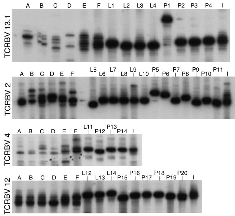
Methods: We used complementarity-determining region 3 (CDR3) length analysis to examine the complete T cell receptor (TCR) beta -chain repertoire in skin-lesion biopsy samples from subjects with genital herpes.
Results: We found that herpetic skin lesions consistently demonstrated oligoclonal CDR3 DNA length distribution, indicating the presence of T cell expansions. Sequence analysis of representative HSV-specific lesional CD4(+) cell clones and TCR beta -variable (TCRBV) sequencing confirmed that the oligoclonal expansions were largely related to HSV-specific T cell proliferation. To assess the persistence of HSV-specific CD4(+) cells that localize to genital lesions, we developed a sensitive and highly specific clonal tracking technique using a combination of TCRBV-specific polymerase chain reaction, followed by liquid hybridization with clonotype-specific probes.
Conclusion: Two different patterns of clonal persistence were observed. Some long-lasting clones appear to home to different epithelia, such as skin and genital mucosa, and to circulate in the peripheral blood, whereas others detected in lesions were absent or very rare in the peripheral blood.
Figures


Similar articles
-
Long term persistence of herpes simplex virus-specific CD8+ CTL in persons with frequently recurring genital herpes.J Immunol. 2000 Jul 15;165(2):1146-52. doi: 10.4049/jimmunol.165.2.1146. J Immunol. 2000. PMID: 10878394
-
Persistence of herpes simplex virus type 2 VP16-specific CD4+ T cells.Hum Immunol. 2005 Jul;66(7):777-87. doi: 10.1016/j.humimm.2005.03.007. Hum Immunol. 2005. PMID: 16112025
-
Homing in on the cellular immune response to HSV-2 in humans.Am J Reprod Immunol. 2005 Apr;53(4):172-81. doi: 10.1111/j.1600-0897.2005.00262.x. Am J Reprod Immunol. 2005. PMID: 15760378 Free PMC article.
-
Immune surveillance by CD8αα+ skin-resident T cells in human herpes virus infection.Nature. 2013 May 23;497(7450):494-7. doi: 10.1038/nature12110. Epub 2013 May 8. Nature. 2013. PMID: 23657257 Free PMC article.
-
Virus-specific immune memory at peripheral sites of herpes simplex virus type 2 (HSV-2) infection in guinea pigs.PLoS One. 2014 Dec 8;9(12):e114652. doi: 10.1371/journal.pone.0114652. eCollection 2014. PLoS One. 2014. PMID: 25485971 Free PMC article.
Cited by
-
Persistence of EBV antigen-specific CD8 T cell clonotypes during homeostatic immune reconstitution in cancer patients.PLoS One. 2013 Oct 25;8(10):e78686. doi: 10.1371/journal.pone.0078686. eCollection 2013. PLoS One. 2013. PMID: 24205294 Free PMC article. Clinical Trial.
-
An extremely diverse CD4 response to vaccinia virus in humans is revealed by proteome-wide T-cell profiling.J Virol. 2008 Jul;82(14):7120-34. doi: 10.1128/JVI.00453-08. Epub 2008 May 14. J Virol. 2008. PMID: 18480455 Free PMC article.
-
High frequency of herpesvirus-specific clonotypes in the human T cell repertoire can remain stable over decades with minimal turnover.J Virol. 2013 Jan;87(1):697-700. doi: 10.1128/JVI.02180-12. Epub 2012 Oct 17. J Virol. 2013. PMID: 23077319 Free PMC article.
-
Persistence of HIV-1 receptor-positive cells after HSV-2 reactivation is a potential mechanism for increased HIV-1 acquisition.Nat Med. 2009 Aug;15(8):886-92. doi: 10.1038/nm.2006. Epub 2009 Aug 2. Nat Med. 2009. PMID: 19648930 Free PMC article. Clinical Trial.
-
Innate immune responses to herpes simplex virus type 2 influence skin homing molecule expression by memory CD4+ lymphocytes.J Virol. 2006 Mar;80(6):2863-72. doi: 10.1128/JVI.80.6.2863-2872.2006. J Virol. 2006. PMID: 16501095 Free PMC article.
References
-
- Koelle DM, Abbo H, Peck A, Ziegweid K, Corey L. Direct recovery of herpes simplex virus (HSV)–specific T lymphocyte clones from recurrent genital HSV-2 lesions. J Infect Dis. 1994;169:956–61. - PubMed
Publication types
MeSH terms
Substances
Grants and funding
LinkOut - more resources
Full Text Sources
Other Literature Sources
Medical
Research Materials
