. 2005 Jun 15;80(6):798-808.
doi: 10.1002/jnr.20474.

Connexin43, the major gap junction protein of astrocytes, is down-regulated in inflamed white matter in an animal model of multiple sclerosis

Affiliations

Connexin43, the major gap junction protein of astrocytes, is down-regulated in inflamed white matter in an animal model of multiple sclerosis

Elimor Brand-Schieber et al. J Neurosci Res. .

Abstract

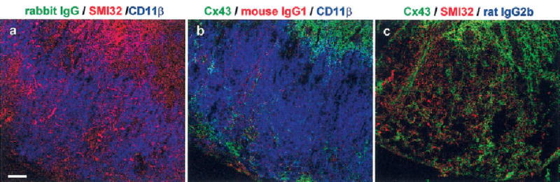
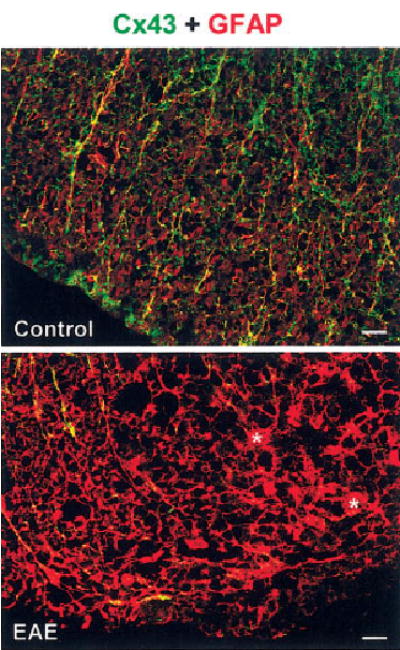
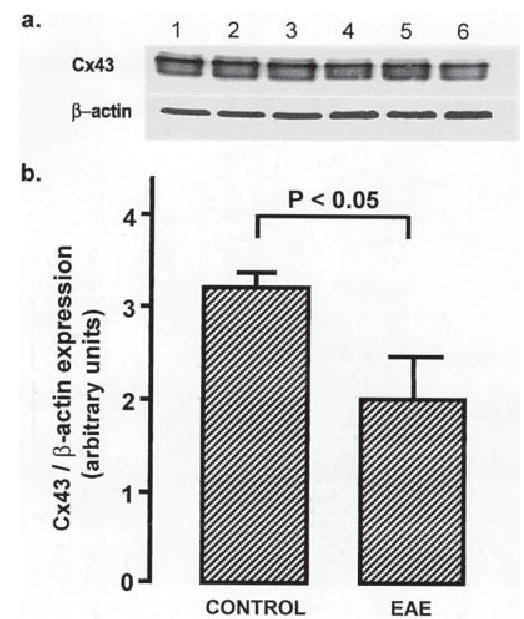
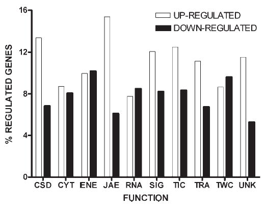
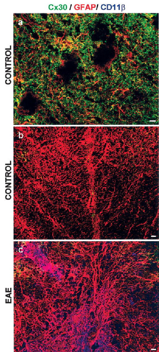
Both multiple sclerosis (MS) and experimental autoimmune encephalomyelitis (EAE), its animal model, involve inflammatory attack on central nervous system (CNS) white matter, leading to demyelination and axonal damage. Changes in astrocytic morphology and function are also prominent features of MS and EAE. Resting astrocytes form a network that is interconnected through gap junctions, composed mainly of connexin43 (Cx43) protein. Although astrocytic gap junctional connectivity is known to be altered in many CNS pathologies, little is known about Cx43 expression in inflammatory demyelinating disease. Therefore, we evaluated the expression of Cx43 in spinal cords of EAE mice compared with healthy controls. Lumbar ventral white matter areas were heavily infiltrated with CD11beta-immunoreactive monocytes, and within these infiltrated regions loss of Cx43 immunoreactivity was evident. These regions also showed axonal dystrophy, demonstrated by the abnormally dephosphorylated heavy-chain neurofilament proteins. Astrocytes in these Cx43-depleted lesions were strongly glial fibrillary acidic protein reactive. Significant loss (38%) of Cx43 protein in EAE mouse at the lumbar portion of spinal cords was confirmed by Western blot analysis. Decreased Cx43 transcript level was also observed on cDNA microarray analysis. In addition to changes in Cx43 expression, numerous other genes were altered, including those encoding adhesion and extracellular matrix proteins. Our data support the notion that, in addition to damage of myelinating glia, altered astrocyte connectivity is a prominent feature of inflammatory demyelination.


Figures

Fig. 1
Fig. 1
Regions showing reduced Cx43 in spinal cords from mice with EAE correlate with monocytes and dystrophic axons. Confocal microscopic images depicting transverse lumbar spinal cord sections from controls and two mice with EAE that were triply labeled for Cx43 (a–c; green), SMI32 (d–f; red), and CD11β (g–i; blue). Merged images are shown in j–l. Control tissue (left row) has intense Cx43 labeling but does not display SMI32 (dystrophic axons) and CD11β (monocytes) labeling. Tissue from animals sick with EAE (middle and right rows) has pockets of reduced Cx43 immunoreactivity. These regions also show increased axonal dystrophy and monocyte infiltration, indicated by enhanced SMI32 and CD11β labeling, respectively. A region clear of CD11β within an inflammatory compartment displays preserved Cx43 labeling (arrow). Scale bar = 50 μm.
Fig. 2
Fig. 2
Control sections show specificity of immunolabeling in spinal cords from mice with EAE. Confocal microscopic images depicting transverse lumbar spinal cord sections from mice with EAE that were labeled by combining all pairs of the following proteins: Cx43 (b,c; green), SMI32 (a,c; red), and CD11β (a,b; blue). In each section, the third primary antibody was replaced with a matching species/IgG subtype antibody obtained from animals that were not exposed to an antigenic peptide. The combinations are specified above each panel. a: No labeling was detected in the green channel when Cx43 antibody was replaced with a nonspecific rabbit IgG. b: Only faint labeling was detected in the red channel when SMI32 antibody was replaced with nonspecific mouse IgG1. c: No labeling was detected in the blue channel when CD11β antibody was replaced with a non-specific rat IgG2b. Scale bar = 50 μm.
Fig. 3
Fig. 3
Regions showing reduced Cx43 in spinal cords from mice with EAE also display hypertrophic astrocytes. Confocal microscopic images depicting transverse lumbar spinal cord section from control (top) and EAE (bottom) that were doubly labeled for Cx43 (green) and the astrocytic marker GFAP (red). Overlap of green and red appears yellow. Control white matter shows Cx43 overlapping with typical GFAP-positive astrocytes. In EAE, regions of reduced Cx43 immunoreactivity contain intensely labeled GFAP-positive astrocytes that appear hypertrophied, with enlarged cell bodies (asterisks) and thick processes. Scale bars = 20 μm.
Fig. 4
Fig. 4
Cx43 protein is reduced in spinal cords of mice with EAE. a: A Western blot demonstrating reduced Cx43 protein in lumbar spinal cord homogenates from mice with EAE (lanes 4–6) compared with controls (lanes 1–3). b: A graph showing semiquantitative analysis based on Cx43 to β-actin ratios (mean ± SEM). There is a significant reduction of Cx43 in EAE (control 3.17 ± 0.12; EAE 1.99 + 0.41; n = 3; P < 0.05, ANOVA followed by Student’s t-test).
Fig. 5
Fig. 5
Functional categories of significantly regulated genes in spinal cords of AT-EAE mice compared to controls. A total of 1,433 out of 7,455 genes with known protein product were significantly (P < 0.05) either up- (open bars) or down-regulated (solid bars) in AT-EAE. In this histogram, numbers of genes regulated within each class are expressed as a percentage of the total number of genes within each class on the array. Genes were categorized as to the following functions: CSD: cell cycle, shape, differentiation, death, CYT: cytoskeleton, ENE: energy and metabolism, JAE: cell junction, adhesion, extracellular matrix, RNA: RNA processing, SIG: cell signaling, TIC, transport of small molecules and ions into the cell, TRA: transcription, TWC: transport of ions/molecules within the cell, UNK: function not yet assigned. Note that the JAE group has the highest percentage of up-regulated (15.4%) and the lowest percentage of down-regulated (6.3%) genes, whereas other gene categories show a more uniform distribution of up- and down-regulation.
Fig. 6
Fig. 6
Cx30 immunoreactivity is not enhanced in inflamed white matter. Confocal microscopic images depicting transverse lumbar spinal cord sections from controls (a,b) and animals with EAE (c) labeled for Cx30 (green), GFAP (red), and CD11β (blue). Gray matter (a) is well labeled for Cx30, which overlaps (appearing yellow) with GFAP. Rounded voids most likely represent motor neurons. There is only little Cx30 immunoreactivity in white matter (b), especially adjacent to gray matter (top edges of the images). No infiltrating cells are apparent as evidenced by the absence of CD11β labeling. In EAE, Cx30 labeling is not enhanced in inflamed monocyte infiltrated white matter (c). Scale bars = 10 μm.

Similar articles

Cited by

References

    1. Adams RD, Kubik CS. The morbid anatomy of the demyelinative disease. Am J Med. 1952;12:510–546. - PubMed
    1. Alexander DB, Goldberg GS. Transfer of biologically important molecules between cells through gap junction channels. Curr Med Chem. 2003;10:2045–2058. - PubMed
    1. Bauer J, Berkenbosch F, Van Dam AM, Dijkstra CD. Demonstration of interleukin-1 beta in Lewis rat brain during experimental allergic encephalomyelitis by immunocytochemistry at the light and ultra-structural level. J Neuroimmunol. 1993;48:13–21. - PubMed
    1. Bauer J, Huitinga I, Zhao W, Lassmann H, Hickey WF, Dijkestra CD. The role of macrophages, perivascular cells, and microglial cells in the pathogenesis of experimental autoimmune encephalomyelitis. Glia. 1995;15:437–446. - PubMed
    1. Benn T, Halfpenny C, Scolding N. Glial cells as targets for cytotoxic immune mediators. Glia. 2001;36:200–211. - PubMed

Publication types

MeSH terms