Comparative Study
. 2005;7(3):R476-84.
doi: 10.1186/ar1694. Epub 2005 Feb 18.

Small GTP-binding protein Rho-mediated signaling promotes proliferation of rheumatoid synovial fibroblasts

Affiliations
Comparative Study

Small GTP-binding protein Rho-mediated signaling promotes proliferation of rheumatoid synovial fibroblasts

Shingo Nakayamada et al. Arthritis Res Ther. 2005.

Abstract

Rho is a major small GTP-binding protein that is involved in the regulation of various cell functions, including proliferation and cell migration, through activation of multiple signaling molecules in various types of cells. We studied its roles in synovial fibroblasts (SFs) in patients with rheumatoid arthritis (RA) and clarified its relevance to RA synovitis, with the following results. 1)We found that the thrombin receptor was overexpressed on RA synovial fibroblasts (RA SFs) and that thrombin induced a marked proliferation and progression of the cell cycle to the S phase in these cells. 2)We also found that thrombin efficiently activated Rho. 3)Rho activation and proliferation and the progression of the cell cycle to the S phase were completely blocked by p115RGS (an N-terminal regulator of the G-protein signaling domain of p115RhoGEF) and by the C-terminal fragments of Galpha13 (an inhibitor of the interaction of receptors with G13). 4)Thrombin induced the secretion of IL-6 by RA SFs, but this action was blocked by p115RGS or Galpha13. Our findings show that the actions of thrombin on the proliferation of RA SFs, cell-cycle progression to the S phase, and IL-6 secretion were mainly mediated by the G13 and RhoGEF pathways. These results suggest that p115RGS and Galpha13 could be potent inhibitors of such functions. A rational design of future therapeutic strategies for RA synovitis could perhaps include the exploitation of the Rho pathway to directly reduce the growth of synovial cells.


Figures

Figure 1
Figure 1
Overexpression of thrombin receptor on SFs from patients with RA. (a) Histograms representing thrombin receptor expression on synovial fibroblasts (SFs) from rheumatoid arthritis (RA) and osteoarthritis (OA) patients. Cells were stained with the antithrombin receptor mAb ATAP2. Flow-cytometric analyses were performed using FACScan. Open histograms represent the number of cells stained with ATAP2 in each logarithmic scale on a fluorescence amplifier. Shaded histograms represent profiles of anti-Thy1.2 mAb as a negative control. (b) Comparison of thrombin receptor expression in RA and OA SFs. The expression of thrombin receptor was analyzed by FACScan. Each value represents the number of molecules expressed per cell, calculated using standard QIFKIT beads from five similar experiments, as described in Materials and methods. Data are expressed as mean ± standard deviation for five independent donors. FACS, fluorescence-activated cell sorter. **P < 0.01.
Figure 2
Figure 2
Thrombin induces synovial proliferation and progression to S phase in RA SFs. Cells were cultured for 48 hours in DMEM containing 1% FCS and then stimulated with the indicated amount of thrombin. For the proliferation assay, at 24 hours after thrombin stimulation, cells were stained with TetraColor One including tetrazolium and electron-carrier mixture for detecting cell proliferation. The optical density (OD) was measured by ELISA plate reader at 450 nm. For analysis of the cell cycle, at 24 hours after thrombin stimulation, cells were collected, washed with PBS, and fixed in 70% ethanol for 2 hours at 4°C. After treatment of cells with 10 μg/ml ribonuclease for 15 min at 37°C, fixed cells were stained with 50 μg/ml propidium iodide for 2 min. The DNA content was subsequently measured by FACScan fluorescence-activated cell sorter. (a) Dose-dependent proliferation of synovial fibroblasts (SFs) from rheumatoid arthritis (RA) patients. The OD was measured by ELISA plate reader at 450 nm. (b) Histogram representing the cell cycle in RA SFs, as detected by FACScan. (c) Dose-dependent S-phase progression of the cell cycle in RA SFs. Numbers represent the percentage of cells exhibiting mean channel fluorescence (FL2-H) in the S/G2/M phase of the cell. Data are expressed as mean ± standard deviation for five experiments, using five independent donors. **P < 0.01 in comparison with the value found without thrombin stimulation.
Figure 3
Figure 3
Effects of thrombin on proliferation and cell cycle in SFs from RA and OA. Cells were cultured for 48 hours in DMEM containing 1% FCS and then stimulated with 10 units/ml thrombin. (a) Time-course of proliferation of synovial fibroblasts (SFs) from patients with rheumatoid arthritis (RA) (dark symbols) and patients with osteoarthritis (OA) (light symbols). Continuous lines, thrombin-stimulated cells; dotted lines, unstimulated cells. The optical density (OD) was measured by ELISA plate reader at 450 nm. (b) Comparison of cell cycle in RA SFs and OA SFs. Numbers represent the percentage of cells exhibiting mean channel fluorescence (FL2-H) in the S/G2/M phase of the cell-division cycle. Data are expressed as mean ± standard deviation of five experiments, using five independent donors. **P < 0.01.
Figure 4
Figure 4
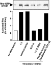
Inhibition of thrombin-induced Rho activation by expression of Gα13-ct and p115RGS in RA synovial fibroblasts. Rheumatoid arthritis (RA) synovial fibroblasts (SFs) were or were not infected with adenoviruses encoding green-fluorescent protein (GFP) (control vector), the C-terminal regions of Gα13 (Gα13-ct), or P115RGS. Cells that were not infected with adenovirus were incubated in the medium alone. Cells were then cultured for 48 hours in DMEM containing 1% FCS, then stimulated with 10 units/ml thrombin for 1 minute or were loaded with GTPγS (positive control), after which they were lysed to measure Rho activity. Rho activity is indicated by the amount of Rho bound by the Rhotekin-Rho-binding domain (RBD) (top). The percentage of activated Rho (graph) is expressed as a ratio relative to 4% of total Rho (4% of total protein used in the RBD bead pull-down experiments). Results are representative of three experiments. Western blot analysis confirmed that equal amounts of total Rho were used for the pull-down assay under each condition (data not shown). (-), cells without infection; p115RGS, regulator of G-protein signaling domain of p115Rho guanine nucleotide exchange factor.
Figure 5
Figure 5
G13 and Rho signaling in thrombin-mediated synovial proliferation and S-phase progression in RA SFs. Rheumatoid arthritis (RA) synovial fibroblasts (SFs) infected or not infected with adenoviruses encoding GFP (control vector), the C-terminal regions of Gα12 (Gα12-ct), Gα13-ct, or p115RGS were cultured for 48 hours in DMEM containing 1% FCS and then stimulated with 10 units/ml thrombin. At 24 hours after the thrombin stimulation, proliferation assay and cell-cycle analysis of RA SFs were performed. (a) Effect of Rho signaling inhibition on thrombin-induced cell proliferation. Numbers represent the optical density (OD) as measured by ELISA plate reader at 450 nm. (b) Effect of Rho signaling inhibition on thrombin-induced cell-cycle progression. Numbers represent the percentage of cells that exhibited mean channel fluorescence (FL2-H) in the S/G2/M phase. Data are expressed as mean ± standard deviation of five experiments, using five independent donors. (-), cells without infection; p115RGS, regulator of G-protein signaling domain of p115Rho guanine nucleotide exchange factor. *P < 0.05, **P < 0.01, in comparison with thrombin stimulation.
Figure 6
Figure 6
Thrombin induces IL-6 secretion via Rho-mediated signaling in RA synovial fibroblasts. Rheumatoid arthritis (RA) synovial fibroblasts (SFs) were or were not infected with adenoviruses encoding green-fluorescent protein (control vector), the C-terminal regions of Gα12 (Gα12-ct), Gα13-ct, or p115RGS. Cells were cultured for 48 hours in DMEM containing 1% FCS, then stimulated with the indicated amount of thrombin. At 3 to 24 hours after thrombin stimulation, the supernatants of cultured cells were collected and assayed for IL-6 using commercial ELISA kits. (a) Dose-dependent IL-6 production by RA SFs at 12 hours after thrombin stimulation. (b) Time course of IL-6 secretion by RA SFs stimulated with 10 units/ml thrombin. (c) Effect of Rho signaling inhibition on thrombin-induced IL-6 secretion at 12 hours after thrombin stimulation. Data are expressed as mean ± standard deviation of five experiments, using five independent donors. (-), cells without infection; p115RGS, regulator of the G-protein signaling domain of p115Rho guanine nucleotide exchange factor. *P < 0.05, **P < 0.01, in comparison with (a) time 0, (b) no thrombin stimulation, and (c) the indicated data.

References

    1. Etienne-Manneville S, Hall A. Rho GTPases in cell biology. Nature. 2002;420:629–635. doi: 10.1038/nature01148. - DOI - PubMed
    1. Bar-Sagi D, Hall A. Ras and Rho GTPases: a family reunion. Cell. 2000;13:227–238. doi: 10.1016/S0092-8674(00)00115-X. - DOI - PubMed
    1. Yamamoto M, Marui N, Sakai T, Morii N, Kozaki S, Ikai K, Imamura S, Narumiya S. ADP-ribosylation of the rhoA gene product by botulinum C3 exoenzyme causes Swiss 3T3 cells to accumulate in the G1 phase of the cell cycle. Oncogene. 1993;8:1449–1455. - PubMed
    1. Rodriguez-Fernandez JL, Sanchez-Martin L, Rey M, Vicente-Manzanares M, Narumiya S, Teixido J, Sanchez-Madrid F, Cabanas C. Rho and Rho-associated kinase modulate the tyrosine kinase PYK2 in T-cells through regulation of the activity of the integrin LFA-1. J Biol Chem. 2001;276:40518–40527. doi: 10.1074/jbc.M102896200. - DOI - PubMed
    1. Van Aelst L, D'Souza-Schorey C. Rho GTPases and signaling networks. Genes Dev. 1997;11:2295–2322. - PubMed

Publication types

MeSH terms