Intra-articular injections of high-molecular-weight hyaluronic acid have biphasic effects on joint inflammation and destruction in rat antigen-induced arthritis
- PMID: 15899053
- PMCID: PMC1174961
- DOI: 10.1186/ar1725
Intra-articular injections of high-molecular-weight hyaluronic acid have biphasic effects on joint inflammation and destruction in rat antigen-induced arthritis
Abstract
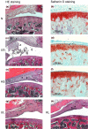
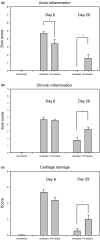
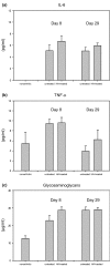
To assess the potential use of hyaluronic acid (HA) as adjuvant therapy in rheumatoid arthritis, the anti-inflammatory and chondroprotective effects of HA were analysed in experimental rat antigen-induced arthritis (AIA). Lewis rats with AIA were subjected to short-term (days 1 and 8, n = 10) or long-term (days 1, 8, 15 and 22, n = 10) intra-articular treatment with microbially manufactured, high-molecular-weight HA (molecular weight, 1.7 x 10(6) Da; 0.5 mg/dose). In both tests, 10 buffer-treated AIA rats served as arthritic controls and six healthy animals served as normal controls. Arthritis was monitored by weekly assessment of joint swelling and histological evaluation in the short-term test (day 8) and in the long-term test (day 29). Safranin O staining was employed to detect proteoglycan loss from the epiphyseal growth plate and the articular cartilage of the arthritic knee joint. Serum levels of IL-6, tumour necrosis factor alpha and glycosaminoglycans were measured by ELISA/kit systems (days 8 and 29). HA treatment did not significantly influence AIA in the short-term test (days 1 and 8) but did suppress early chronic AIA (day 15, P < 0.05); however, HA treatment tended to aggravate chronic AIA in the long-term test (day 29). HA completely prevented proteoglycan loss from the epiphyseal growth plate and articular cartilage on day 8, but induced proteoglycan loss from the epiphyseal growth plate on day 29. Similarly, HA inhibited the histological signs of acute inflammation and cartilage damage in the short-term test, but augmented acute and chronic inflammation as well as cartilage damage in the long-term test. Serum levels of IL-6, tumour necrosis factor alpha, and glycosaminoglycans were not influenced by HA. Local therapeutic effects of HA in AIA are clearly biphasic, with inhibition of inflammation and cartilage damage in the early chronic phase but with promotion of joint swelling, inflammation and cartilage damage in the late chronic phase.
Figures


Similar articles
-
Novel hyaluronic acid-methotrexate conjugate suppresses joint inflammation in the rat knee: efficacy and safety evaluation in two rat arthritis models.Arthritis Res Ther. 2016 Apr 1;18:79. doi: 10.1186/s13075-016-0971-8. Arthritis Res Ther. 2016. PMID: 27039182 Free PMC article.
-
Intraarticular injection of platelet-rich plasma reduces inflammation in a pig model of rheumatoid arthritis of the knee joint.Arthritis Rheum. 2011 Nov;63(11):3344-53. doi: 10.1002/art.30547. Arthritis Rheum. 2011. PMID: 21769848
-
Cartilage-specific constitutive expression of TSG-6 protein (product of tumor necrosis factor alpha-stimulated gene 6) provides a chondroprotective, but not antiinflammatory, effect in antigen-induced arthritis.Arthritis Rheum. 2002 Aug;46(8):2207-18. doi: 10.1002/art.10555. Arthritis Rheum. 2002. PMID: 12209527
-
Beneficial actions of hyaluronan (HA) on arthritic joints: effects of molecular weight of HA on elasticity of cartilage matrix.Biorheology. 2006;43(3,4):347-54. Biorheology. 2006. PMID: 16912407 Review.
-
Hyaluronic acid, an efficient biomacromolecule for treatment of inflammatory skin and joint diseases: A review of recent developments and critical appraisal of preclinical and clinical investigations.Int J Biol Macromol. 2018 Sep;116:572-584. doi: 10.1016/j.ijbiomac.2018.05.068. Epub 2018 May 29. Int J Biol Macromol. 2018. PMID: 29772338 Review.
Cited by
-
Effects of hyaluronan treatment on lipopolysaccharide-challenged fibroblast-like synovial cells.Arthritis Res Ther. 2007;9(1):R1. doi: 10.1186/ar2104. Arthritis Res Ther. 2007. PMID: 17214881 Free PMC article.
-
Extracellular matrix-blood composite injection reduces post-traumatic osteoarthritis after anterior cruciate ligament injury in the rat.J Orthop Res. 2016 Jun;34(6):995-1003. doi: 10.1002/jor.23117. Epub 2015 Dec 18. J Orthop Res. 2016. PMID: 26629963 Free PMC article.
-
Efficacy of an immunotoxin to folate receptor beta in the intra-articular treatment of antigen-induced arthritis.Arthritis Res Ther. 2012 May 2;14(3):R106. doi: 10.1186/ar3831. Arthritis Res Ther. 2012. PMID: 22551402 Free PMC article.
-
Aspects of the biology of hyaluronan, a largely neglected but extremely versatile molecule.Wien Med Wochenschr. 2006 Nov;156(21-22):563-8. doi: 10.1007/s10354-006-0351-0. Wien Med Wochenschr. 2006. PMID: 17160372 Review.
-
A Network Pharmacology Approach to Explore Mechanism of Action of Longzuan Tongbi Formula on Rheumatoid Arthritis.Evid Based Complement Alternat Med. 2019 Jan 17;2019:5191362. doi: 10.1155/2019/5191362. eCollection 2019. Evid Based Complement Alternat Med. 2019. PMID: 30792744 Free PMC article.
References
-
- Firestein GS, Zvaifler NJ. Rheumatoid arthritis: a disease of disordered immunity. In: Gallin J, Goldstein I, Willis K, editor. Inflammation: Basic Principles and Clincal Correlates. New York: Raven Press; 1992. pp. 959–975.
-
- Mau W, Bornmann M, Weber H, Weidemann HF, Hecker H, Raspe HH. Prediction of permanent work disability in a follow-up study of early rheumatoid arthritis: results of a tree structured analysis using RECPAM. Br J Rheumatol. 1996;35:652–659. - PubMed
-
- Laurent TC, Laurent UB, Fraser JR. The structure and function of hyaluronan: an overview. Immunol Cell Biol. 1996;74:1–7. - PubMed
Publication types
MeSH terms
Substances
LinkOut - more resources
Full Text Sources
Other Literature Sources

