. 2005 May 15;65(10):4191-201.
doi: 10.1158/0008-5472.CAN-04-3865.

Distinct structural domains within C19ORF5 support association with stabilized microtubules and mitochondrial aggregation and genome destruction

Affiliations

Distinct structural domains within C19ORF5 support association with stabilized microtubules and mitochondrial aggregation and genome destruction

Leyuan Liu et al. Cancer Res. .

Abstract

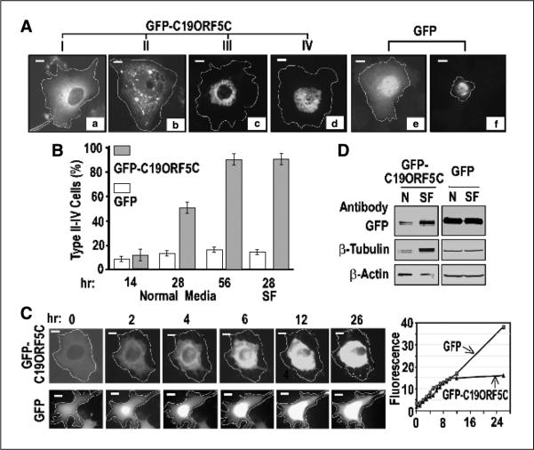
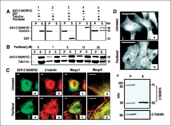
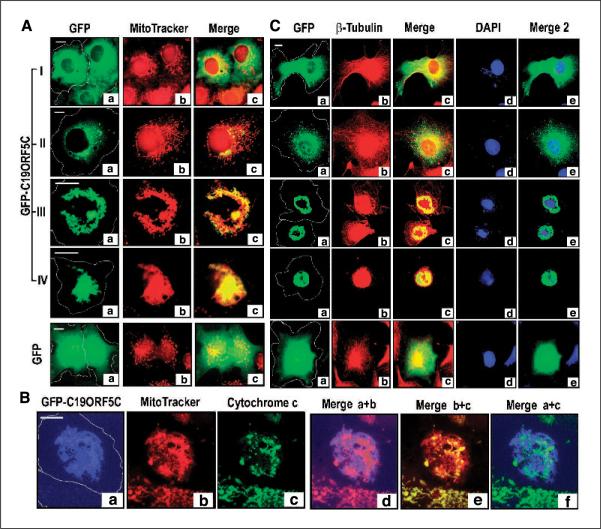
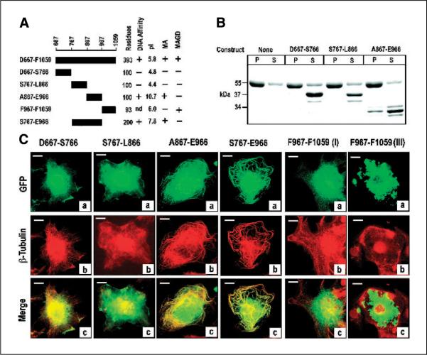
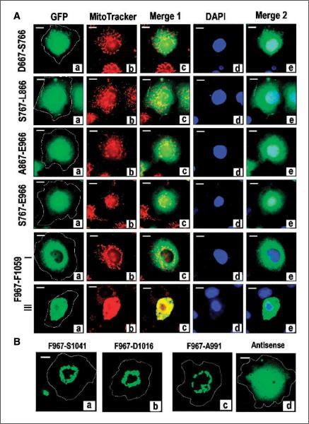
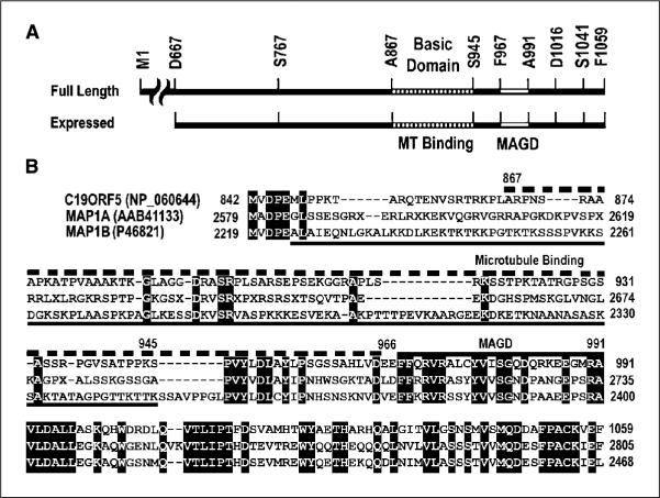
C19ORF5 is a sequence homologue of microtubule-associated proteins MAP1A/MAP1B of unknown function, except for its association with mitochondria-associated proteins and the paclitaxel-like microtubule stabilizer and candidate tumor suppressor RASSF1A. Here, we show that when overexpressed in mammalian cells the recombinant 393-amino acid residue COOH terminus of C19ORF5 (C19ORF5C) exhibited four types of distribution patterns proportional to expression level. Although normally distributed throughout the cytosol without microtubular association, C19ORF5C specifically accumulated on stabilized microtubules in paclitaxel-treated cells and interacted directly with paclitaxel-stabilized microtubules in vitro. The native 113-kDa full-length C19ORF5 and a shorter 56-kDa form similarly associated with stabilized microtubules in liver cells and stabilized microtubules from their lysates. As C19ORF5 accumulated, it appeared on mitochondria and progressively induced distinct perinuclear aggregates of mitochondria. C19ORF5 overlapped with cytochrome c-deficient mitochondria with reduced membrane potential. Mitochondrial aggregation resulted in gross degradation of DNA, a cell death-related process we refer to as mitochondrial aggregation and genome destruction (MAGD). Deletion mutagenesis revealed that the C19ORF5 hyperstabilized microtubule-binding domain resides in a highly basic sequence of <100 residues, whereas the MAGD activity resides further downstream in a distinct 25-residue sequence (F967-A991). Our results suggest that C19ORF5 mediates communication between the microtubular cytoskeleton and mitochondria in control of cell death and defective genome destruction through distinct bifunctional structural domains. The accumulation of C19ORF5 and resultant MAGD signaled by hyperstabilized microtubules may be involved in the tumor suppression activity of RASSF1A, a natural microtubule stabilizer and interaction partner with C19ORF5, and the taxoid drug family.


Figures

Figure 1
Figure 1
Cellular distribution patterns of GFP-C19ORF5C. A, four general morphologic profiles of GFP distribution in COS7 cells transfected with GFP-C19ORF5C. Cells were classified as type I to IV according to increasing association with punctiform aggregates. Cell boundaries were located by light microscope and then determined by increasing exposure times to maximum to detect total GFP signal across the field. The transfected cell periphery was then outlined as indicated. Field exposure was then reduced to facilitate maximum resolution of the high-intensity punctate structures. Bars, 10 μm in all photomicrographs. B, time-dependent increase in type II to IV cells exhibiting punctiform aggregates of GFP-C19ORF5C. Columns, mean counts of type I to IV cell types from three independent experiments in which at least 500 total transfected cells among 50 microscopic fields were counted at the indicated times; bars, SD. Normal medium contained 5% fetal bovine serum. SF, serum-free medium. The number of cells exhibiting the conventional apoptotic morphology in (A, f) was scored in cultures transfected with GFP alone (open columns). C, time-dependent perinuclear clustering of GFP-C19ORF5C in single transfected cells at room temperature. Images were captured at the indicated times from single cells observed continuously. Representative cell of replicate observations and time-dependent increase in intensity of GFP fluorescence in single cells expressing GFP-C19ORF5C or GFP. Relative florescence intensity was determined by dividing the average fluorescence intensity in arbitrary units in a field with brightest florescence in the cells by that of a field of the same size outside the cell boundary. D, direct analysis of total GFP-C19ORF5 or GFP, β-tubulin, and β-actin in transfected cells. GFP-C19ORF5C or GFP transfected COS7 cells were cultured in 25 cm2 tissue culture flasks in medium with (N) or without 5% fetal bovine serum (SF) for 28 hours. Extracts were made from 8 × 10⁵ cells collected by scraping into 200 μL buffer I. An equal amount of soluble protein (160 μg) was applied to each lane of SDS-PAGE, and after electrophoretic transfer, GFP-C19ORF5C, GFP, tubulins, and actin were detected with 1 μg/mL polyclonal antibody against GFP or monoclonal antibodies against β-tubulin or β-actin. Bands were visualized with 0.1 βg/mL alkaline phosphatase–conjugated anti-rabbit IgG or anti-mouse IgG antibodies.
Figure 2
Figure 2
Specific association of the C19ORF5 with paclitaxel-stabilized microtubules. A, association of purified GST-C19ORF5C with paclitaxel-stabilized microtubules. Tubulins (10 μg) were incubated with 10 μg GST-C19ORF5C, GST, or other control GST fusion proteins in G-PEM buffer for 30 minutes at 37°C in the presence of 5 μmol/L paclitaxel. The 20 μL mixtures were layered on 10 μL of 10% sucrose solution in PEM buffer and centrifuged at 10,000 × g for 20 minutes at room temperature. Supernatant (S, 30 μL) containing soluble tubulins and other relatively low molecular weight components and pellet (P) resuspended in 30 μL G-PEM buffer containing stabilized microtubules and associated proteins were loaded side by side on 7.5% SDS-PAGE. Protein bands were visualized with Coomassie blue stain. B, paclitaxel concentration-dependent association of GST-C19ORF5C protein with microtubules. Microtubules were assembled in the presence of the same amount of GST-C19ORF5C and tubulin employed in (A) with the indicated amounts of paclitaxel. C, specific association of the C19ORF5 with paclitaxel-stabilized microtubules in vivo. COS7 cells treated with 10 μmol/L paclitaxel where indicated at the time of transfection with cDNA coding for GFP-C19ORF5C were stained with anti-β-tubulin and Texas red–conjugated anti-mouse antibody. The relationship of GFP (green) and anti-β-tubulin antibody (red) fluorescence was observed and colors merged to test for overlap of signals (yellow). Merge2, segments of cells in Merge1 magnified 2.5 times. Representative of 90% of transfected cells from three independent experiments. D, distribution of native C19ORF5 in normal (a) and paclitaxel-treated HepG2 cells (b) and its association with paclitaxel-stabilized microtubules from HepG2 cell lysate in vitro (c). Cells were treated with paclitaxel as indicated and analyzed with mAb4G1. Representative of the majority of cells in three independent experiments. For in vitro assembly of microtubules, ~1 × 10⁶ HepG2 cells were lysed in 150 μL buffer and followed by sonication for 10 seconds. The cell lysate was clarified by centrifugation at 10,000 × g for 20 minutes and treated with 10 μmol/L paclitaxel at 37°C for 1 hour to stabilize microtubules and the reaction mixture was loaded on a cushion of 20 μL of 20% sucrose. Stabilized microtubules and associated proteins were sedimented by ultracentrifugation (Sorvall RC M120) at 100,000 × g for 1 hour. The pellet containing the stabilized microtubules was resuspended in 150 μL buffer. An equal volume of pellet and supernatant (25 μL) fractions were analyzed on SDS-PAGE and immunoblotted with antibodies against C19ORF5 (mAb4G1) or β-tubulin, respectively. FL, full-length C19ORF5 predicted from cDNA; SC, short chain.
Figure 3
Figure 3
MAGD caused by C19ORF5. A, relationship of GFP-C19ORF5C and mitochondria in type I to IV cells. Cell boundaries were determined and then outlined as described in Fig. 1. The green GFP signal (a) was merged with that of red MitoTracker (b) in Merge (c). Yellow, overlap of the GFP signal with mitochondria. B, relation of GFP-C19ORF5C-associated mitochondria with cytochrome c. To achieve maximum color contrast, the strong GFP signal from GFP-C19ORF5C was artificially assigned blue. The cytochrome c signal was assigned green. The overlap of blue GFP-C19ORF5C with red MitoTracker, red MitoTracker with green cytochrome c, or blue GFP-C19ORF5C with green cytochrome c is indicated by purple, yellow, or cyan in Merge d, e, or f, respectively. C, coincidence of microtubular collapse and DNA degradation with C19ORFC-associated mitochondrial aggregation. The green GFP signal from GFP-C19ORF5 (a) was analyzed with that of red β-tubulin immunochemical stain (b) in Merge (c) and blue DAPI DNA stain (d) in Merge 2 (e). Yellow in Merge is indicative of overlap of the GFP signal with tubulin and cyan in Merge 2 is indicative of overlap of the GFP signal with DAPI. Microtubules were visualized by immunoanalysis as described in Materials and Methods. Control cells transfected with GFP alone are indicated.
Figure 4
Figure 4
Identification of the hyperstabilized microtubule-binding domain within the C19ORF5 COOH terminus in vitro. A, sequence and properties of constructs. The indicated constructs were fused with GST or GFP at their NH2 terminus. The GST fusions were expressed, recovered, and purified using glutathione and DNA affinity as described in Materials and Methods. Numbering is according to full-length cDNA of C19ORF5, single letter code for amino acids at the NH2- and COOH-terminal residue of the product, and the total residues are indicated. Physicochemical and functional properties of the construct products are summarized on the right. Theoretical pI values were calculated using the Web-based tool from http://us.expasy.org/tools/pi_tool.html. MA, microtubule association activity; MAGD, cytotoxic MAGD activity; nd, not determined. B, binding to paclitaxel-stabilized microtubules in vitro. Association of the indicated constructs with paclitaxel-stabilized microtubules assembled from tubulins was determined as described in Fig. 1A. The 55-kDa band is tubulin and the 37-kDa or smaller bands are the GST fusions and their lower molecular weight truncates. As indicated in Materials and Methods, purified products of the A867-E966 construct were ≤34 kDa. Insufficient recovery of purified GST-tagged F967-F1059 from bacteria prevented assessment in vitro. The purified GST-S767-E966 with predicted molecular weight of 47 kDa overlapped tubulin on SDS-PAGE and is not shown. P, pellet containing microtubules; S, supernatant containing unpolymerized tubulin. C, confirmation of the hyperstabilized microtubule-binding domain in C19ORF5. COS cells transfected with the indicated constructs with GFP at the NH2 terminus were treated with paclitaxel and cells were examined for association of the green GFP signal with hyperstabilized microtubules as well as overlap with red anti-β-tubulin as in Fig. 2. Representative of near 100% of transfected cells with the first four constructs, 90% and 10% of transfected cells with F967-F1059, respectively, in at least three independent experiments in which >2,000 cells were examined.
Figure 5
Figure 5
Identification of an independent MAGD domain. A, analysis of GFP-tagged constructs coding for subdomains of C19ORF5C. COS cells were transfected with the indicated constructs with GFP at the NH2 terminus and cells were stained with MitoTracker and DAPI dyes. Cultures were examined for type I to IV phenotypes 24 hours after transfection in the same way as described for GFP-C19ORF5C. Yellow in Merge 1 and cyan in Merge 2 indicate overlap of green GFP-C19ORF5C with mitochondria (red) or DNA (blue), respectively. Representative cell from each transfected culture. Type II to IV cell morphologies were only observed in cultures transfected with F967-F1059 at the relative frequencies described for GFP-C19ORF5C in Fig. 1. Representative type I and III cell. B, reduction of the MAGD domain to a 25-residue sequence. The indicated constructs of MAGD domain-containing GFP-F967-F1059 with sequential deletions at the COOH terminus were tested for induction of the type II to IV phenotypes indicative of MAGD activity. Antisense, coding sequence for GFP fused to the in-frame antisense coding sequence for F967-A991.
Figure 6
Figure 6
Comparison of functional sequence domains in the COOH terminus of C19ORF5 to sequence of the light chains of MAP1A and MAP1B. A, sequence domain structure of C19ORF5. Full-length C19ORF5 and the 393–amino acid residue C19ORF5C (D667-F1059) with residues flanking constructs used in this study are indicated. A867-S945 indicates the highly basic microtubule-binding domain. F967-A991 is the MAGD domain. B, sequence alignment of human C19ORF5 with the light chains of MAP1A (MAP1A-LC2) and MAP1B (MAP1B-LC1). Identical residues in all three sequences are in black. Dashed bar (top), C19ORF5 sequence A867-E966 containing the microtubule-binding domain; bottom solid line, microtubule-binding domain of MAP1B (45); top solid bar, MAGD domain of C19ORF5.

Similar articles

Cited by

References

    1. Lengauer C, Kinzler KW, Vogelstein B. Genetic instabilities in human cancers. Nature. 1998;396:643–9. - PubMed
    1. Duesberg P, Rasnick D. Aneuploidy, the somatic mutation that makes cancer a species of its own. Cell Motil Cytoskeleton. 2000;47:81–107. - PubMed
    1. Jallepalli PV, Lengauer C. Chromosome segregation and cancer: cutting through the mystery. Nat Rev Cancer. 2001;1:109–17. - PubMed
    1. Schneider BL, Kulesz-Martin M. Destructive cycles: the role of genomic instability and adaptation in carcino-genesis. Carcinogenesis. 2004;25:2033–44. - PubMed
    1. Sorger PK, Dobles M, Tournebize R, Hyman AA. Coupling cell division and cell death to microtubule dynamics. Curr Opin Cell Biol. 1997;9:807–14. - PubMed

Publication types

MeSH terms