. 2005 Jun;25(11):4359-70.
doi: 10.1128/MCB.25.11.4359-4370.2005.

Trm11p and Trm112p are both required for the formation of 2-methylguanosine at position 10 in yeast tRNA

Affiliations

Trm11p and Trm112p are both required for the formation of 2-methylguanosine at position 10 in yeast tRNA

Suresh K Purushothaman et al. Mol Cell Biol. 2005 Jun.

Abstract

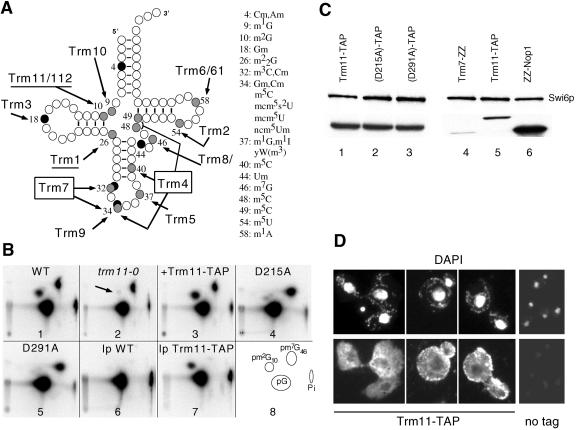
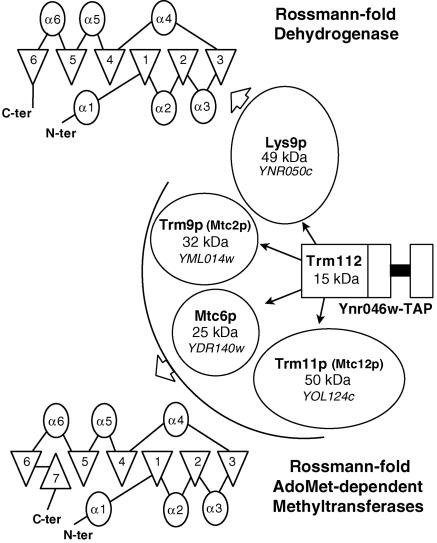
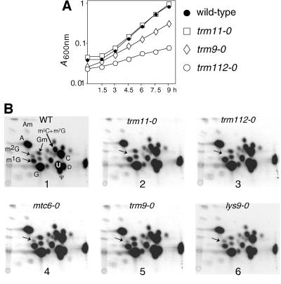
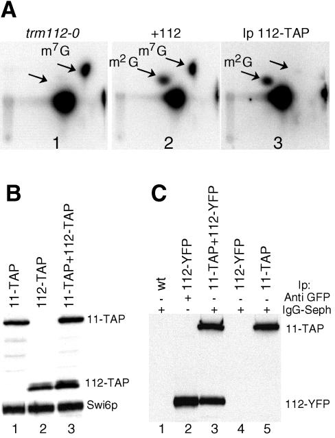
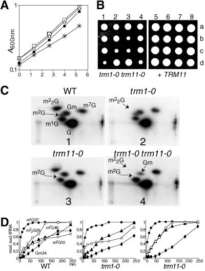
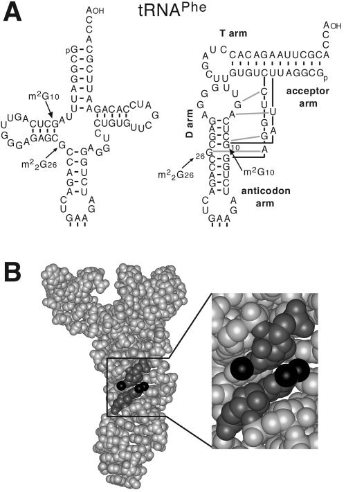
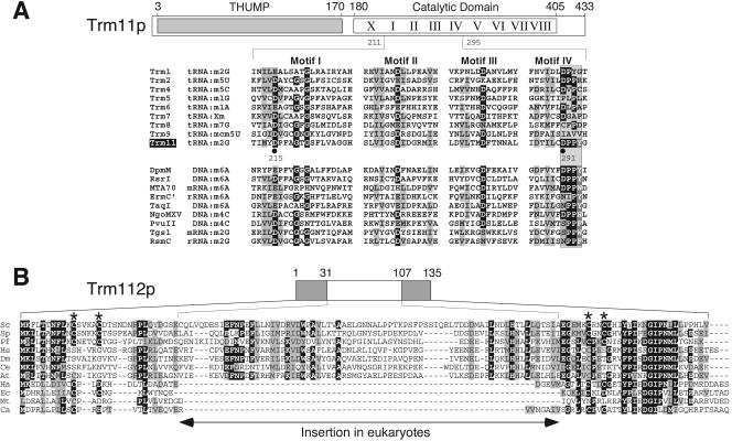
N(2)-Monomethylguanosine-10 (m(2)G10) and N(2),N(2)-dimethylguanosine-26 (m(2)(2)G26) are the only two guanosine modifications that have been detected in tRNA from nearly all archaea and eukaryotes but not in bacteria. In Saccharomyces cerevisiae, formation of m(2)(2)G26 is catalyzed by Trm1p, and we report here the identification of the enzymatic activity that catalyzes the formation of m(2)G10 in yeast tRNA. It is composed of at least two subunits that are associated in vivo: Trm11p (Yol124c), which is the catalytic subunit, and Trm112p (Ynr046w), a putative zinc-binding protein. While deletion of TRM11 has no detectable phenotype under laboratory conditions, deletion of TRM112 leads to a severe growth defect, suggesting that it has additional functions in the cell. Indeed, Trm112p is associated with at least four proteins: two tRNA methyltransferases (Trm9p and Trm11p), one putative protein methyltransferase (Mtc6p/Ydr140w), and one protein with a Rossmann fold dehydrogenase domain (Lys9p/Ynr050c). In addition, TRM11 interacts genetically with TRM1, thus suggesting that the absence of m(2)G10 and m(2)(2)G26 affects tRNA metabolism or functioning.


Figures

FIG. 1.
FIG. 1.
Trm11p is required for m2G10 formation in vitro. (A) Cloverleaf representation of yeast tRNA showing the position of methylated nucleotides. Black circles, 2′-O-methylriboses; shaded circles, methylated bases. The position and the nature of the modified nucleotides are listed on the right side of the figure. The 11 known tRNA MTase activities (including Trm11/Trm112) are shown, with their target(s). The sites of action of the two multisite specific enzymes, Trm4 and Trm7, have been boxed. (B) In vitro methylation analysis of [α-32P]GTP-radiolabeled intronless tRNAIleUAU, using S10 extracts prepared from various strains. Panels: 1, wild-type (BMA64); 2, trm11-0 (YBL4577); 3, trm11-0 plus Trm11-TAP (YBL4588); 4, Trm11-D215A-TAP mutant protein (YBL4597); 5, Trm11-D291A-TAP mutant protein (YBL4598); 6, control immunoprecipitation using a wild-type extract and IgG-Sepharose beads; 7, immunoprecipitation of Trm11-TAP (YBL4580); 8, reference map indicating the location of the three nucleotides of interest (pG, pm2G10, and pm7G46). Pi, inorganic phosphate. The arrow points to the m2G10 spot that is absent in the trm11-0 strain. The solvent system used for the TLC was NI/RII (37). (C) Western blot analysis of TAP-tagged proteins. Similar amounts of protein were loaded in each lane, as demonstrated by the detection of Swi6p (47), which is used here as a control. Comparison of the signal obtained for wild-type Trm11p (lane 1) and for the two mutant proteins D215A (lane 2) and D291A (lane 3) is shown. The signal obtained for Trm11-TAPp (lane 5) was compared with those obtained for Trm7-ZZp (lane 4) (51) and ZZ-Nop1p (lane 6) (21). (D) Immunofluorescence detection of Trm11-TAP in yeast cells. (Top) Cells were labeled with DAPI to visualize the structures containing DNA, nuclei and mitochondria (different fields with representative cells are shown). (Bottom) Fluorescent IgGs detect the S. aureus protein A fragment, expressed as a fusion with Trm11p. No tag, wild-type cells expressing no tagged protein (lower magnification).
FIG. 2.
FIG. 2.
Trm112p interacts with several proteins, including Trm11p. Large-scale analysis of protein complexes (22) has revealed an interaction of Trm112p with Lys9p, Trm9p, Mtc6p, and Trm11p, as schematically shown. Molecular masses of the five proteins are given in kilodaltons. A secondary structure representation is shown for the Rossmann fold and for the MTase domain. α-Helices are represented by ovals and β-strands are represented by triangles.
FIG. 3.
FIG. 3.
Trm11p and Trm112p are both required in vivo for the formation of m2G10. (A) Growth curves of wild-type and mutant strains in YPD at 30°C. Black circles, wild type (BMA64); open squares, trm11-0 (YBL4577); open diamonds, trm9-0 (YBL4557); open circles, trm112-0 (YBL4663). A600 nm was plotted on a semilogarithmic graph as a function of time in hours. (B) Autoradiogram of selected 2D-TLC of modified nucleotides after nuclease P1 digestion of in vivo-labeled [32P]tRNA. Hydrolysates of total tRNA were analyzed with the chromatographic system NI/RII (37). Panels: 1, wild type (WT); 2, trm11-0; 3, trm112-0; 4, mtc6-0; 5, trm9-0; 6, lys9-0. The spots of interest are shown on the wild-type panel. The arrows indicate the positions for m2G10.
FIG. 4.
FIG. 4.
Trm112p is required for the formation of m2G10 in vitro and is associated with Trm11p. (A) [α-32P]GTP-labeled intronless tRNAIleUAU was incubated with S10 cell extracts prepared from various strains or with an immunoprecipitated fraction, and then modified nucleotides were analyzed as in Fig. 1. Panel 1, trm112-0 (YBL4663); panel 2, +112 (strain trm112-0 complemented with a TRM112 gene on a centromeric plasmid [YBL4665]); panel 3, Ip 112-TAP (Trm112-TAPp immunoprecipitated from an S100 extract prepared from strain YBL4634 and tested for m2G10 formation activity). (B) Comparison of the abundance of Trm112-TAPp and Trm11-TAPp. Western blot analysis was performed using IgGs coupled to peroxidase, and extracts were prepared from cells expressing protein A-tagged proteins. Lanes: 1, Trm11-TAPp(YBL4580); 2, Trm112-TAPp(YBL4634); 3, Trm11-TAPp/Trm112-TAPp(YBL4635). Similar amounts of proteins were loaded in each lane, as demonstrated by using anti-Swi6p antibodies. (C) Coimmunoprecipitation experiment. Cellular extracts prepared from strains expressing either Trm11-TAPp, Trm112-YFPp, or both were incubated with IgG-Sepharose beads, the pellets were washed under stringent conditions, and then proteins were eluted and tested by Western blot analysis, using an anti-GFP serum that detects both fusion proteins. Lane 1, wild-type strain; lanes 2 and 4, Ynr046w-YFP; lane 3, YBL4689; lane 5, YBL4580. In the results shown in lane 2, immunoprecipitation was performed using anti-GFP and protein A-Sepharose beads.
FIG. 5.
FIG. 5.
TRM11 and TRM112 interact genetically with TRM1. (A) Growth curves of various strains at 30°C in YPD. Filled circles, wild-type; open triangles, trm1-0; open squares, trm11-0; asterisks, trm1-0 trm11-0. (B) Spore analysis of a diploid strain heterozygous for the two loci trm1-0::URA3/TRM1 and trm11-0::kanMX4/TRM11 (YBL4611) or the same strain transformed with a plasmid containing the TRM11 wild-type gene and an LEU2 marker. Spores 1d, 2c, 3b, 3c, and 4d were trm1-0 trm11-0. Spores 5a, 5b, 6a, 7d, and 8b were also deleted for trm1-0 and trm11-0; however, they contained the TRM11 gene on an LEU2 plasmid (pBL640). Each colony was scored for Ura+, Leu+, or G418 resistance on appropriate panels. (C) 2D-TLC analysis of [α-32P]GTP-labeled intronless tRNAPhe incubated with S10 cell extracts as shown in Fig. 1. Panels: 1, extracts prepared from a wild-type strain (WT); 2, trm1-0; 3, trm11-0; 4, the double-mutant strain trm1-0 trm11-0. Arrows point to the spots corresponding to m2G10, m22G26, and Gm34. (D) Kinetic analysis of the formation of modified nucleotides in vitro, using S10 extracts prepared from wild-type (WT), trm1-0, and trm11-0 strains. For each time point, nucleotides were separated by 2D-TLC, and each spot was quantified by phosphorimaging.
FIG. 6.
FIG. 6.
(A) Cloverleaf and L-shape representations (35) of yeast tRNAPhe, depicting the relative positions of m2G10 and m22G26. (B) Three-dimensional structure of tRNAPhe (1eHz file, available at http://www.resb.org/pdb/) with the two methylguanosines stacked on each other and a close-up view indicating the direction of the two methyl groups.
FIG. 7.
FIG. 7.
Sequence and structure analysis of Trm11p and Trm112p. (A) A schematic representation of Trm11p, with its two structural domains: the N-terminal THUMP (residues 1 to 180, shaded box) and the C-terminal catalytic RFM (with nine conserved motifs indicated by roman numerals). The sequence of Trm11p from S. cerevisiae is shown for a region corresponding to the most conserved motifs I to IV (residues 211 to 295), of which the AdoMet-binding motifs I to III are common to other tRNA MTases from yeast (top), but only the catalytic motif IV is evidently common with representative exocyclic amino MTases (bottom). The catalytic DPPY motif has been boxed. Identical and physicochemically similar residues are shown on a black and gray background, respectively. The two critical aspartate residues (D215 and D291) that have been mutated are shown with a black dot. (B) The sequence alignment of Trm112p from S. cerevisiae (Sc) and its orthologs from six eukaryotes (Sp, Schizosaccharomyces pombe; Pf, Plasmodium falciparum; Hs, Homo sapiens; Dm, Drosophila melanogaster; Ce, Caenorhabditis elegans; At, Arabidopsis thaliana), one archaeon (Ha; Halobacterium sp. NRC-1), and three bacteria (Ec, E. coli; Mt, Mycobacterium tuberculosis; Ca, C. aurantiacus).

References

    1. Alexandrov, A., M. R. Martzen, and E. M. Phizicky. 2002. Two proteins that form a complex are required for 7-methylguanosine modification of yeast tRNA. RNA 8:1253-1266. - PMC - PubMed
    1. Anderson, J., L. Phan, R. Cuesta, B. A. Carlson, M. Pak, K. Asano, G. R. Björk, M. Tamame, and A. G. Hinnebusch. 1998. The essential Gcd10p-Gcd14p nuclear complex is required for 1-methyladenosine modification and maturation of initiator methionyl-tRNA. Genes Dev. 12:3650-3662. - PMC - PubMed
    1. Aravind, L., and E. V. Koonin. 2001. THUMP: a predicted RNA-binding domain shared by 4-thiouridine, pseudouridine synthases and RNA methylases. Trends Biochem. Sci. 26:215-217. - PubMed
    1. Arluison, V., C. Hountondji, B. Robert, and H. Grosjean. 1998. Transfer RNA-pseudouridine synthetase Pus1 of Saccharomyces cerevisiae contains one atom of zinc essential for its native conformation and tRNA recognition. Biochemistry 37:7268-7276. - PubMed
    1. Armengaud, J., J. Urbonavicius, B. Fernandez, G. Chaussinand, J. M. Bujnicki, and H. Grosjean. 2004. N2-Methylation of guanosine at position 10 in tRNA is catalyzed by a THUMP domain-containing, S-adenosylmethionine-dependent methyltransferase, conserved in Archaea and Eukaryota. J. Biol. Chem. 279:37142-37152. - PubMed

Publication types

MeSH terms