. 2005 Jun;25(11):4488-500.
doi: 10.1128/MCB.25.11.4488-4500.2005.

Gadd45a expression induces Bim dissociation from the cytoskeleton and translocation to mitochondria

Affiliations

Gadd45a expression induces Bim dissociation from the cytoskeleton and translocation to mitochondria

Tong Tong et al. Mol Cell Biol. 2005 Jun.

Abstract

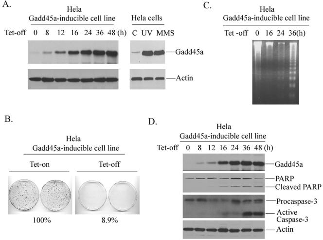
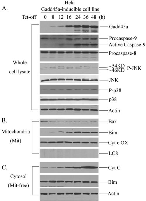
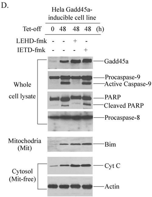
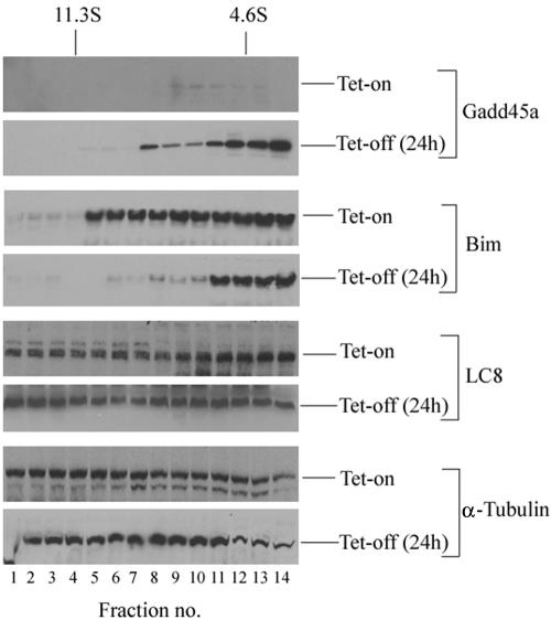
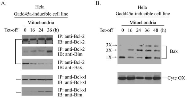
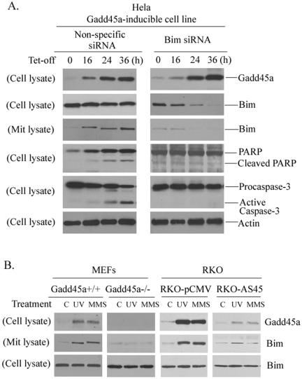
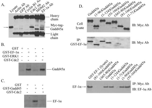
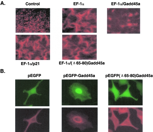
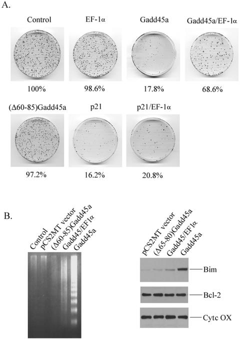
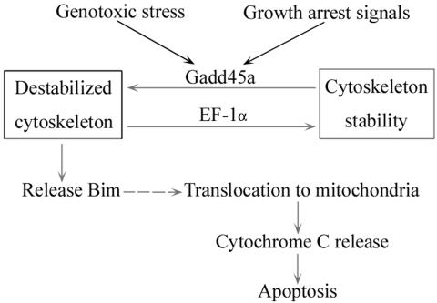
Gadd45a, a p53- and BRCA1-regulated stress protein, has been implicated in the maintenance of genomic fidelity, probably through its roles in the control of cell cycle checkpoint and apoptosis. However, the mechanism(s) by which Gadd45a is involved in the induction of apoptosis remains unclear. We show here that inducible expression of Gadd45a protein causes dissociation of Bim, a Bcl2 family member, from microtubule-associated components and translocation to mitochondria. The Bim accumulation in mitochondria enhances interaction of Bim with Bcl-2, relieves Bax from Bcl-2-bound complexes, and subsequently results in release of cytochrome c into the cytoplasm. Suppression of endogenous Bim greatly inhibits Gadd45a induction of apoptosis. Interestingly, Gadd45a interacts with elongation factor 1alpha (EF-1alpha), a microtubule-severing protein that plays an important role in maintaining cytoskeletal stability, and inhibits EF-1alpha-mediated microtubule bundling, indicating that the interaction of Gadd45a with EF-1alpha disrupts cytoskeletal stability. A mutant form of Gadd45a harboring a deletion of EF-1alpha-binding domain fails to inhibit microtubule stability and to induce Bim translocation to mitochondria. Furthermore, coexpression of EF-1alpha antagonizes Gadd45a's property of suppressing cell growth and inducing apoptosis. These findings identify a novel link that connects stress protein Gadd45a to the apoptotic machinery and address the importance of cytoskeletal stability in apoptotic response to DNA damage.


Figures

FIG. 1.
FIG. 1.
Inducible expression of Gadd45a results in apoptosis in HeLa cells. (A) a HeLa Gadd45a-inducible cell line was established as described in Materials and Methods. Cells were placed in 100-mm dishes at a density of 4 × 10⁵ and grown in DMEM containing tetracycline at a concentration of 2 μg/ml. After withdrawal of the tetracycline, the cells were collected at the indicated time points for preparation of cellular protein. A total of 100 μg of whole-cell protein was used for immunoblotting analysis with anti-Gadd45a antibody. In addition, Gadd45a protein levels in HeLa cells treated with UV at a dose of 10 J/m2 or MMS at a concentration of 100 μg/ml were examined. As a loading control, anti-actin antibody was included. (B) HeLa Gadd45a-inducible cells were seeded at a density of 1,000 cells per 100-mm dish and grown in medium containing 2 μg of tetracycline/ml. After 16 h, the medium was removed, the plates were washed three times with PBS, and then fresh medium containing no tetracycline was added to the plates. The cells were fixed and stained at 14 days and scored for colonies containing at least 50 cells. The experiments were performed four times, and only representative results are shown here. (C and D) After inducible expression of Gadd45a protein, cells were collected at the indicated time points for the assay of cellular DNA fragmentation (C) or for measurements of cleavages of PARP and caspase-3 (D).
FIG. 2.
FIG. 2.
Bim induction in mitochondria by Gadd45a protein. (A) The HeLa Gadd45a-inducible cell line was grown in DMEM in the presence of tetracycline at a concentration of 2 μg/ml. After removal of the tetracycline, the cells were collected at the indicated time points for the preparation of cellular protein. A total of 100 μg of whole-cell protein was examined for activation of caspase-9, caspase-8, JNK, and p38 kinases. In addition, total JNK and p38 protein were also examined. (B) Mitochondrial protein was isolated from HeLa Gadd45a-inducible cells (see Materials and Methods) at different time points and used for the detection of Bim and Bax. (C) Cytochrome c release was measured in mitochondrion (Mit)-free cytosol protein after inducible expression of Gadd45a in Hthe removal of tetracycline, caspase-9 inhibitor (LEHD-fmk) or caspase-8 inhibitor (IETD-fmk) was added to the cell cultures. After 48 h, the cells were collected for the preparation of cellular protein. A total of 100 μg of whole-cell protein was examined for the activation of caspase-9, caspase-8, and PARP cleavage. Bim induction of mitochondria and cytochrome c release were examined as in panels B and C.
FIG. 2.
FIG. 2.
Bim induction in mitochondria by Gadd45a protein. (A) The HeLa Gadd45a-inducible cell line was grown in DMEM in the presence of tetracycline at a concentration of 2 μg/ml. After removal of the tetracycline, the cells were collected at the indicated time points for the preparation of cellular protein. A total of 100 μg of whole-cell protein was examined for activation of caspase-9, caspase-8, JNK, and p38 kinases. In addition, total JNK and p38 protein were also examined. (B) Mitochondrial protein was isolated from HeLa Gadd45a-inducible cells (see Materials and Methods) at different time points and used for the detection of Bim and Bax. (C) Cytochrome c release was measured in mitochondrion (Mit)-free cytosol protein after inducible expression of Gadd45a in Hthe removal of tetracycline, caspase-9 inhibitor (LEHD-fmk) or caspase-8 inhibitor (IETD-fmk) was added to the cell cultures. After 48 h, the cells were collected for the preparation of cellular protein. A total of 100 μg of whole-cell protein was examined for the activation of caspase-9, caspase-8, and PARP cleavage. Bim induction of mitochondria and cytochrome c release were examined as in panels B and C.
FIG. 3.
FIG. 3.
Inducible expression of Gadd45a causes dissociation of Bim from cytoskeleton. HeLa Gadd45a-inducible cells were cultured in DMEM containing tetracycline at a concentration of 2 μg/ml. At 24 h after withdrawal of the tetracycline, cells were treated with 5 μM Taxol (Sigma) for 4 h and 0.5 mM AMP-PNP for 30 min before lysis. Cellular protein extracts were prepared and fractionated by sucrose gradient sedimentation (see Materials and Methods). Proteins in gradient fractions were size separated by gel electrophoresis, transferred to nitrocellulose membranes, and immunoblotted with antibodies to Gadd45a, Bim, LC8, and β-tubulin.
FIG. 4.
FIG. 4.
Mitochondrion accumulation of Bim after Gadd45a induction increases physical association of Bim with Bcl-2 and enhances Bax multimerization. (A) After inducible expression of Gadd45a protein in the HeLa cell line, cells were collected at the indicated time points. Mitochondrial lysates were prepared (see Materials and Methods) to examine the interactions between Bim and Bcl-2 or Bcl-XL. Lysates were incubated with 10 μl of antibody to Bcl-2 or Bcl-XL and 20 μl of protein A/G agarose beads (Santa Cruz Biotechnology, Santa Cruz, CA) at 4°C for 4 h. Immunocomplexes were washed four times with lysis buffer and examined by immunoblot analysis for the existence of Bim and Bax. (B) After Gadd45a induction, cells were collected at the indicated time points and subjected to treatment with 1 mM DSP (cross-linking agent) at 37°C for 30 min before subcellular fractionation. Mitochondrial proteins were analyzed with antibodies to Bax.
FIG. 5.
FIG. 5.
Suppression of endogenous Bim expression inhibits Gadd45a-induced apoptosis. (A) A total of 5 × 10⁵ HeLa Gadd45a-inducible cells were plated into 100-cm dishes and grown in DMEM containing tetracycline. Upon removal of the tetracycline, the cells were transfected with 21-nucleotide siRNA duplexes that target Bim mRNA transcript via oligofectamine (see Materials and Methods). As a control, nonspecific 21-nucleotide siRNA duplexes were included in the experiments. Cells were collected at the indicated times for preparation of whole-cell lysates or mitochondrial protein. The protein levels of Bim in both whole lysates and mitochondria were analyzed by Western immunoblot assays. In addition, cleavages of PARP and caspase-3 were examined in cells treated with Bim siRNA. (B) Detection of Bim mitochondrial induction in cells with disrupted Gadd45a. MEFs derived from Gadd45a knockouts and RKO-AS45 that expresses antisense Gadd45a were treated with UV radiation at a dose of 10 J/m2 or MMS at a concentration of 50 μg/ml. After 12 h, cells were harvested for the preparation of both whole-cell lysates and mitochondrial protein. Immunoblotting assays were performed to examine the Bim protein levels of mitochondria.
FIG. 6.
FIG. 6.
Interaction of Gadd45a with EF-1α. (A) Myc-tagged-Gadd45a vector was transiently expressed in HeLa cells via Lipofectamine transfection. At 48 h posttransfection, whole-cell protein extracts were prepared and immunoprecipitated with anti-actin, anti-GFP, anti-Cdc2, anti-MDM2, and anti-EF-1α antibodies. The immunocomplexes were analyzed by sodium dodecyl sulfate-polyacrylamide gel electrophoresis and Western blotting with anti-c-Myc antibody. (B) Whole-cell lysates prepared from HeLa cells were incubated with GST-EF-1α, GST-ERK1, GST-Cdc2, or GST alone (conjugated with Sepharose beads) for 6 h at 4°C. Immunocomplexes were then washed four times with lysis buffer and analyzed with antibody to Gadd45a. (C) GST-Gadd45a, GST-Cdc2, and GST alone were incubated with HeLa cellular protein as described in panel B. Immunoblotting assays were used to examine the presence of EF-1α protein in immunocomplexes by using anti-EF-1α antibody. (D) A series of Myc-tagged-Gadd45a deletion mutants were introduced into HeLa cells via transient transfection. Whole-cell lysates were prepared and incubated with GST-EF-1α (conjugated with Sepharose beads). Immunocomplexes were then washed four times with lysis buffer and analyzed with anti-Myc antibody. In addition, cellular lysates isolated from HeLa cells transfected with a series of Myc-tagged Gadd45a deletion mutants were incubated with GST-EF-1α protein and immunoprecipitated with anti-Myc antibody. Immunoprecipitates were analyzed with anti-EF-1α antibody.
FIG. 7.
FIG. 7.
(A) Effect of Gadd45a on EF-1α-mediated microtubule bundling. Microtubule assembly and/or bundle assays were carried out by using rhodamine (or fluorescein)-labeled tubulin according to the manufacturer's manual. The assays were performed in the presence of 100 ng of GST-EF-1α, GST-Gadd45a, or GST-p21 proteins and evaluated by using fluorescence microscopy. (B) Effect of Gadd45a expression on cytoskeleton. GFP-Gadd45a and GFP(Δ65-80)Gadd45a expression vectors were transfected into HeLa cells. After 36 h, cells were stained with antibody to β-tubulin and detected by fluorescence microscopy for green fluorescent protein (green fluorescence) or rhodamine staining (red fluorescence).
FIG. 8.
FIG. 8.
Interaction of Gadd45a with EF-1α contributes to Gadd45a-induced apoptosis and growth suppression. (A) HeLa cells were cotransfected with pCS2MT (Myc-tagged empty vector) or the indicated Myc-tagged expression vectors for Gadd45a, (Δ60-85)Gadd45a, EF-1α, or p21waf1/cip1. After selection with G418 for 2 weeks, cells were fixed, and the colonies that contained at least 50 cells were counted. Quantitative results represent the average of three individual experiments. (B) HeLa cells were transiently transfected with Myc-tagged Gadd45a or Myc-tagged (Δ60-85)Gadd45a. (C) At 48 h posttransfection, cells were harvested for the assay of DNA fragmentation and for analysis of Bim induction in mitochondria.
FIG. 9.
FIG. 9.
Schematic representation of signaling pathway that mediates Gadd45a-induced apoptosis. In cellular response to genotoxic stress and growth arrest signals, Gadd45a is highly induced. Increased Gadd45a protein interacts with EF-1α, a microtubule-severing protein that maintains cytoskeletal stability, and disrupts the stability of the cytoskeleton. Bim protein is in turn released from destabilized microtubule-associated protein complexes and translocates to mitochondria. Induction of Bim in mitochondria relieves Bax from Bcl-2-bound complexes and induces release of cytochrome c. Finally, cells undergo apoptosis.

References

    1. Adams, J. M., and S. Cory. 2001. Life-or-death decisions by the Bcl-2 protein family. Trends Biochem. Sci. 26:61-66. - PubMed
    1. Antonsson, B., and J. C. Martinou. 2000. The Bcl-2 protein family. Exp. Cell Res. 256:50-57. - PubMed
    1. Carrier, F., M. L. Smith, I. Bae, K. E. Kilpatrick, T. J. Lansing, C. Y. Chen, M. Engelstein, S. H. Friend, W. D. Henner, T. M. Gilmer, et al. 1994. Characterization of human Gadd45, a p53-regulated protein. J. Biol. Chem. 269:32672-32677. - PubMed
    1. Edmonds, B. T., J. Wyckoff, Y. G. Yeung, Y. Wang, E. R. Stanley, J. Jones, J. Segall, and J. Condeelis. 1996. Elongation factor-1 alpha is an overexpressed actin binding protein in metastatic rat mammary adenocarcinoma. J. Cell Science 109:2705-2714. - PubMed
    1. Fan, W., S. Jin, T. Tong, H. Zhao, F. Fan, M. J. Antinore, B. Rajasekaran, M. Wu, and Q. Zhan. 2002. BRCA1 regulates GADD45 through its interactions with the OCT-1 and CAAT motifs. J. Biol. Chem. 277:8061-8067. - PubMed

Publication types

MeSH terms