. 2005 Jun;25(11):4591-601.
doi: 10.1128/MCB.25.11.4591-4601.2005.

Bim regulation of lumen formation in cultured mammary epithelial acini is targeted by oncogenes

Affiliations

Bim regulation of lumen formation in cultured mammary epithelial acini is targeted by oncogenes

Mauricio J Reginato et al. Mol Cell Biol. 2005 Jun.

Abstract

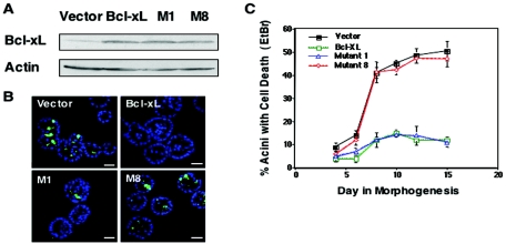
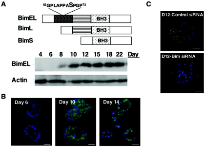
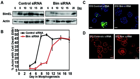
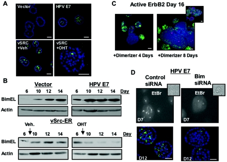
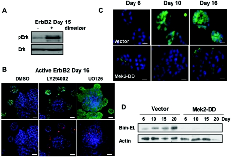
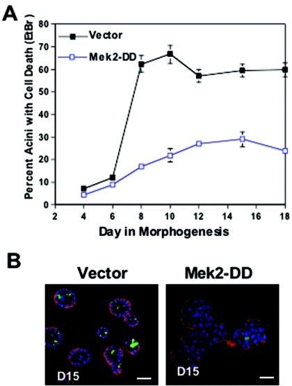
Epithelial cells organize into cyst-like structures that contain a spherical monolayer of cells that enclose a central lumen. Using a three-dimensional basement membrane culture model in which mammary epithelial cells form hollow, acinus-like structures, we previously demonstrated that lumen formation is achieved, in part, through apoptosis of centrally localized cells. We demonstrate that the proapoptotic protein Bim may selectively trigger apoptosis of the centrally localized acinar cells, leading to temporally controlled lumen formation. Bim is not detectable during early stages of three-dimensional mammary acinar morphogenesis and is then highly upregulated in all cells of acini, coincident with detection of apoptosis in the centrally localized acinar cells. Inhibition of Bim expression by RNA interference transiently blocks luminal apoptosis and delays lumen formation. Oncogenes that induce acinar luminal filling, such as ErbB2 and v-Src, suppress expression of Bim through a pathway dependent on Erk-mitogen-activated protein kinase; however, HPV 16 E7, an oncogene that stimulates cell proliferation but not luminal filling, is unable to reduce Bim expression. Thus, Bim is a critical regulator of luminal apoptosis during mammary acinar morphogenesis in vitro and may be an important target of oncogenes that disrupt glandular epithelial architecture.


Figures

FIG. 1.
FIG. 1.
Bcl-xL mutants implicate BH3-only proteins in regulating luminal apoptosis during morphogenesis. (A) Lysates from MCF-10A cells infected with retrovirus of empty vector (MIG), Bcl-xL, Bcl-xL (M1), or Bcl-xL (M8) were analyzed for Bcl-xL, and actin levels were analyzed by Western analysis. (B) At day 10 of morphogenesis, MCF-10A cells expressing vector, Bcl-xL, Bcl-xL (M1), or Bcl-xL (M8) were fixed and immunostained with anti-active caspase-3 (green), and nuclei were counterstained with TOPRO-3 (blue). Scale bar, 20 μm. (C) The percentage of acini containing at least two EtBr-positive cells was measured at the indicated time points during morphogenesis. Values represent the mean ± the SEM of three independent experiments. At least 100 acini were counted in each experiment.
FIG. 2.
FIG. 2.
The BH3-only protein BimEL is upregulated during morphogenesis. (A) Schematic of the three most common Bim splice variants: BimEL, BimL, and BimS. All three isoforms contain a BH3 domain, but only BimEL contains consensus Erk site serine 69. Lysates were prepared from acini at the indicated days in morphogenesis and analyzed by Western analysis with antibodies to Bim and actin. (B) Cryosections (7 μm) of acini from days 6, 10, and 14 of morphogenesis were immunostained with antibody to Bim (green) and counterstained with DAPI (blue). Scale bar, 10 μm. (C) At 24 h after transfection with Bim siRNA oligonucleotides or control oligonucleotides, cells were placed in morphogenesis assays and then fixed, cryosectioned, and immunostained with an antibody to Bim (green) and counterstained with DAPI (blue) at day 12. Scale bar, 10 μm.
FIG. 3.
FIG. 3.
Bim expression involved in luminal apoptosis and luminal clearance during morphogenesis. (A) At 24 h after transfection with Bim siRNA oligonucleotides or matched control oligonucleotides, cells were placed in morphogenesis assays, and cell lysates were collected at the indicated times. Protein levels were analyzed by immunoblotting with Bim or actin antibodies. (B) Cells transfected with Bim or matched control siRNA oligonucleotides were placed in a morphogenesis assay, and the percentage of acini with EtBr-positive cells (two or more cells) was scored after the indicated number of days in culture. Values represent the mean ± the SEM of four independent experiments. (C) Cells transfected with Bim or matched control siRNA oligonucleotides were placed in morphogenesis and were fixed and immunostained at day 10 with anti-active caspase-3 (green), and nuclei were counterstained with TOPRO-3 (blue). Scale bar, 20 μm. (D) Nuclei of representative day 12 structures from cells transfected with Bim or matched control siRNA oligonucleotides were stained with TOPRO-3 (red). Scale bar, 20 μm.
FIG. 4.
FIG. 4.
Phosphorylation of Bim by matrix signals provides protection from Bim's apoptotic function. (A) Lysates were prepared from acini at indicated days in morphogenesis and analyzed by Western analysis with antibodies to pS69-Bim, Bim, and actin. The nonspecific doublet recognized by the pS69-Bim antibody is indicated by an asterisk. (B) Stable Bcl-2 expressing MCF-10A cells were infected with vector (LPCX), BimEL, or BimEL-SA. At 48 h postinfection, cells were treated with trypsin and either replated or placed in suspension. After 24 h the lysates were collected, the proteins were separated by gel electrophoresis, and the samples were immunoblotted with antibodies for pS69-Bim, Bim, or actin. A nonspecific band recognized by the pS69-Bim antibody is indicated by an asterisk. (C) Cells were infected with an equal titer of vector (LPCX), BimEL, or BimEL-SA retrovirus. Bcl-xL-expressing cells were used to determined the titers of BimEl and BimEl-SA virus. The T126G variant BimEL cDNA that is unable to be spliced to form BimL was used for these studies. At 48 h postinfection cells were collected and then analyzed for apoptosis by DNA fragmentation ELISA. Values represent the mean and the standard deviation of A405 to A490 for at least three independent experiments.
FIG. 5.
FIG. 5.
Luminal filling of acini by oncogenes involves downregulation of Bim. (A) Acini expressing vector (LXSN) (day 12), HPV E7 (day 12), or v-Src-ER (treated on day 8 with vehicle control [ethanol] or OHT [1 μM] and analyzed after 48 h) were fixed and immunostained with antibodies to activated caspase-3 (green). Nuclei were counterstained with TOPRO-3 (blue). (B) Lysates from acini expressing vector control or HPV E7 and acini expressing v-Src-ER, treated with either vehicle control (ethanol) or OHT (1 μM) at day 7, were collected at the indicated times. Proteins were separated by gel electrophoresis, and samples were immunoblotted with antibodies for Bim or actin. (C) Cells expressing ErbB2 were treated on day 8 with AP1510 for four (left panel) or eight (right panel) days and were immunostained with antibodies to activated caspase-3 (red) or Bim (green). Nuclei were counterstained with TOPRO-3 (blue). The inset image shows multiacinar ErbB2 structures costained with antibodies to Ki67 (red), integrin-α6 (green), or TOPRO-3 (blue) to demonstrate the accessibility of structures to antibodies. (D) Cells expressing HPV E7 were placed in morphogenesis assays 24 h after transfection with Bim or matched control siRNA oligonucleotides and treated at day 7 with EtBr (top panel; with a corresponding phase image [inset]) or immunostained (bottom panel) at day 12 with an antibody to activated caspase-3 (green), and nuclei were counterstained with TOPRO-3 (blue). Scale bar, 20 μm.
FIG. 6.
FIG. 6.
Involvement of the Mek/Erk pathway in regulating Bim expression. (A) Cells expressing ErbB2 were incubated at day 8 with vehicle control (ethanol) or dimerizer (AP1510), lysates were collected at day 15, and proteins were analyzed by immunoblotting with antibodies to phospho-Erk and Erk. (B) Cells expressing ErbB2 were incubated with AP1510 at day 8 for 8 days and consequently treated with dimethyl sulfoxide, LY294002 (50 μM), or UO126 (40 μM) for 48 h, and D18 acini were immunostained with antibodies to Bim (green; top panel) and activated caspase-3 (red; bottom panel). Nuclei were counterstained with TOPRO-3. Scale bar, 20 μm. (C) Cells expressing vector (Babe) or Mek2-DD were placed in a morphogenesis assay and immunostained at the indicated times with DAPI (blue) and antibody to Bim (green). Scale bar, 10 μm. (D) Cells expressing vector or Mek2-DD were placed in a morphogenesis assay, lysates were collected at the indicated times, and proteins were analyzed by immunoblotting with antibodies to Bim or actin.
FIG. 7.
FIG. 7.
The Mek/Erk pathway regulates luminal apoptosis and luminal filling. (A) Cells expressing vector (Babe) and Mek2-DD were placed in morphogenesis assays, and apoptosis was measured by counting the EtBr-positive acini as described above. Values represent the mean ± the SEM of three independent experiments. (B) Cells expressing vector and Mek2-DD were placed in a morphogenesis assay and immunostained at day 12 with TOPRO-3 (blue), activated caspase-3 (green), and integrin-α6 (red). Scale bar, 20 μm.

Similar articles

Cited by

References

    1. Biswas, S. C., and L. A. Greene. 2002. NGF down-regulates the BH3-only protein Bim and suppresses its proapoptotic activity by phosphorylation. J. Biol. Chem. 277:49511-49516. - PubMed
    1. Blatchford, D. R., L. H. Quarrie, E. Tonner, C. McCarthy, D. J. Flint, and C. J. Wilde. 1999. Influence of microenvironment on mammary epithelial cell survival in primary culture. J. Cell Physiol. 181:304-311. - PubMed
    1. Bouillet, P., S. Cory, L. C. Zhang, A. Strasser, and J. M. Adams. 2001. Degenerative disorders caused by Bcl-2 deficiency prevented by loss of its BH3-only antagonist Bim. Dev. Cell 1:645-653. - PubMed
    1. Cheng, E. H., M. C. Wei, S. Weiler, R. A. Flavell, T. W. Mak, T. Lindsten, and S. J. Korsmeyer. 2001. BCL-2, BCL-X(L) sequester BH3 domain-only molecules preventing BAX- and BAK-mediated mitochondrial apoptosis. Mol. Cell 8:705-711. - PubMed
    1. Coucouvanis, E., and G. R. Martin. 1999. BMP signaling plays a role in visceral endoderm differentiation and cavitation in the early mouse embryo. Development 126:535-546. - PubMed

Publication types

MeSH terms