. 2005 Jun;25(11):4683-92.
doi: 10.1128/MCB.25.11.4683-4692.2005.

Smad1 and Smad8 function similarly in mammalian central nervous system development

Affiliations

Smad1 and Smad8 function similarly in mammalian central nervous system development

Mark Hester et al. Mol Cell Biol. 2005 Jun.

Abstract

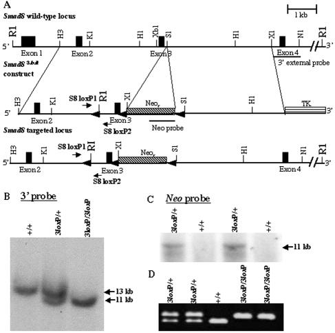
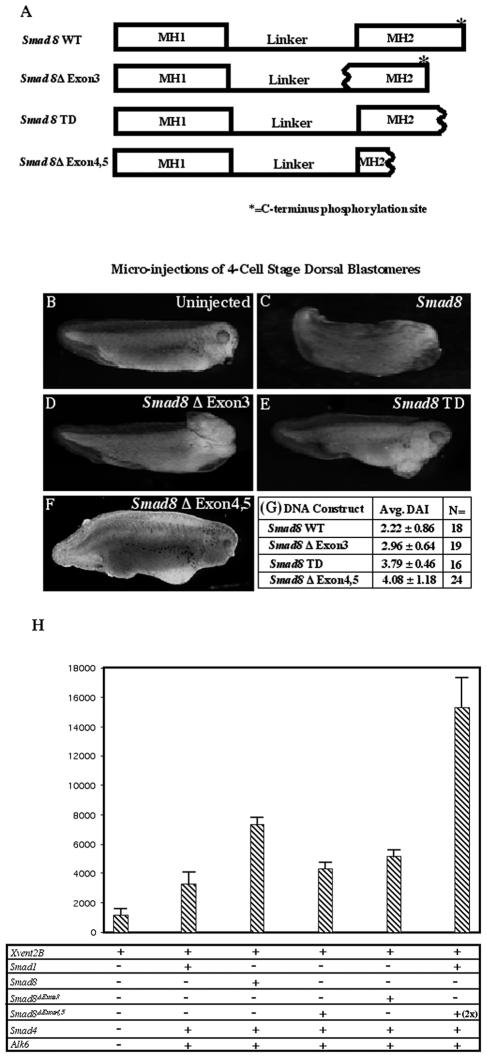
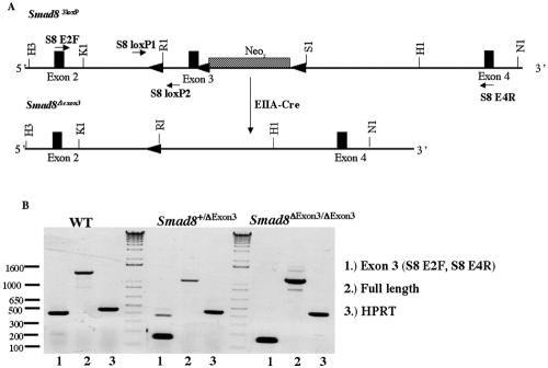
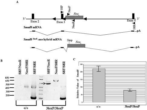
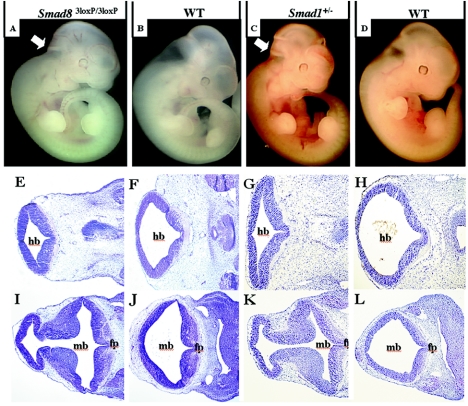
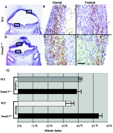
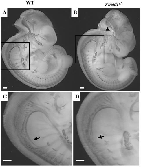
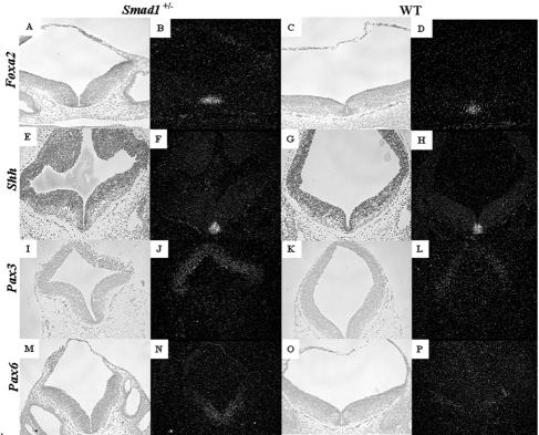
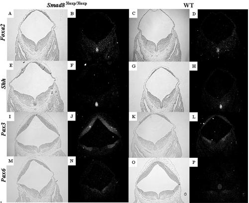
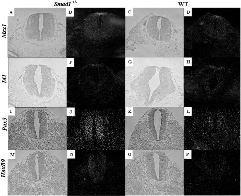
Smads 1, 5, and 8 are the intracellular mediators for the bone morphogenetic proteins (BMPs), which play crucial roles during mammalian development. Previous research has shown that Smad1 is important in the formation of the allantois, while Smad5 has been shown to be critical in the process of angiogenesis. To further analyze the BMP-responsive Smads, we disrupted the murine Smad8 gene utilizing the Cre/loxP system. A Smad8 hypomorphic allele (Smad8(Deltaexon3)) was constructed that contains an in-frame deletion of exon 3, removing one-third of the MH2 domain and a small portion of the linker region. Xenopus injection assays indicated that this Smad8 deletion allele is still functional but has reduced ventralizing capability compared to the wild type. Although Smad8(Deltaexon3/Deltaexon3) embryos are phenotypically normal, homozygotes of another hypomorphic allele of Smad8 (Smad8(3loxP)) containing a neomycin cassette within intron 3, phenocopy an embryonic brain defect observed in roughly 22% of Smad1(+/)(-) embryos analyzed at embryonic day 11.5. These observations suggest that BMP-responsive Smads have critical functions in the development of the mammalian central nervous system.


Figures

FIG. 1.
FIG. 1.
Targeting the murine Smad8 gene. (A) Partial map of the Smad8 genomic locus containing four of the five exons (top), the Smad83loxP targeting vector (middle), and the targeted Smad8 locus (bottom). (B) Southern blot analysis of tail biopsy DNA confirming correct gene targeting of Smad8. Targeting of the Smad8 locus yields a wild-type 13-kb band and a targeted 11-kb band upon digestion of genomic DNA with EcoRI and hybridization with the 3′ external probe indicated in panel A. (C) Further Southern blot analysis with the same digestion scheme using a neomycin probe confirmed correct targeting. An 11-kb band is detected with a neomycin-specific probe. (D) PCR analysis of tail biopsy DNA extracted from Smad83loxP/3loxP mice. Primers were designed within intron 2 flanking the inserted loxP site (A). The lower and upper bands represent the targeted and wild-type alleles, respectively. R1, EcoRI; H3, HindIII; K1, KpnI; H1, HpaI; Xb1, XbaI; S1, SalI; X1, XhoI; N1, NdeI.
FIG. 2.
FIG. 2.
The Smad8Δexon3 allele acts as a hypomorph in Xenopus laevis. (A) Schematic representations of Smad8 WT, Smad8Δexon3, Smad8Δexon4,5, and Smad8 TD constructs used for microinjections into four-cell-stage embryos. Wavy lines represent deletion breakpoints. Smad8Δexon3 lacks one-third of the MH2 domain and a small portion of the linker region. Smad8Δexon4,5 lacks the majority of the MH2 domain. Smad8 TD lacks the C-terminal phosphorylation site. (B) Uninjected embryos develop normally. (C) Microinjection of Smad8 WT mRNA into dorsal blastomeres ventralizes Xenopus tadpoles. (D) Microinjection of Smad8Δexon3 into dorsal blastomeres also ventralizes Xenopus tadpoles but to an intermediate extent. (E) Smad8 TD lacks the majority of the Smad8 ventralizing capability. (F) Microinjection of Smad8Δexon4,5 into dorsal blastomeres also ventralizes Xenopus tadpoles but to a lesser extent. Note, β-galactosidase mRNA was coinjected with Smad8Δexon4,5 mRNA in the image shown in panel F. (G) Average (Avg.) DAI scores and numbers of embryos (N) areshown. (H) Results from a luciferase reporter assay utilizing the Xvent2B promoter driving luciferase and cDNAs for the following constructs: Smad1, Smad8, Smad4, Alk-6, Smad8Δexon3, and Smad8Δexon4,5.
FIG. 3.
FIG. 3.
Cre mediates the excision of exon 3 and neo in Smad83loxP/3loxP mice. (A) Schematic diagram of the Smad83loxP allele undergoing the Cre-mediated deletion of neo and exon 3 from the Smad8 locus. (B) RT-PCR analysis illustrating that exon 3 has been excised from Smad8 in Smad8Δexon3exon3 mice. Lanes 1 show PCR products amplified by primers (S8 E2F and S8 E4R) that flank exon 3 and give a product of ∼400 bp. The 150-bp band seen in the Smad8Δexon3/+ and Smad8Δexon3exon3 samples represent the regions that flank exon 3. Lanes 2 show PCR products of full-length Smad8. As a positive control, hypoxanthine phosphoribosyltransferase (HPRT) was used and is shown in lanes 3. Restriction enzyme abbreviations are given in the legend to Fig. 1.
FIG. 4.
FIG. 4.
Aberrant splicing of neo within Smad8 results in a Smad8-neo fusion transcript. (A) Schematic diagram of the Smad83loxP allele (top) with primer pairs represented as triangles within exons (S8F and S8R) or in the neo cassette (NeoF and NeoR). The Smad8 transcript is depicted (middle) showing exons 2, 3, and 4. The Smad8-neo hybrid transcript found in the Smad83loxP/3loxP mice is spliced in between exons 3 and 4, causing neo to be out of frame with the Smad8 transcript, which results in a premature stop codon within neo. (B) RT-PCR analysis of the Smad8-neo hybrid transcript, with primer pairs shown on top and the indicated sample cDNA shown below. The PCR product using primer pair S8F-NeoR in the Smad83loxP/3loxP sample indicated by an arrow was sequenced and shown to be a Smad8-neo hybrid transcript. (C) Relative level of Smad8 expression as determined by real-time PCR.
FIG. 5.
FIG. 5.
Phenotypic Smad83loxP/3loxP and Smad1+/ embryos display reductions in the hindbrain and midbrain. Genotypes of pictured embryos and sections are shown at the top. (A to D) Whole mount pictures of phenotypic Smad83loxP/3loxP(A), littermate control (B), phenotypic Smad1+/ (C), and littermate control (D) embryos at E11.5. Note that the hindbrain and midbrain are significantly reduced both in phenotypic Smad83loxP/3loxP and Smad1+/ embryos as indicated by arrows. (E to L) Frontal histological sections within the neural tube show reduced space and hypercellularity within the midbrain of phenotypic Smad83loxP/3loxP (I) and phenotypic Smad1+/ (K) embryos at E11.5 compared to wild-type siblings (J and L, respectively). More caudal histological sections show the same diminished space within the hindbrain of abnormal Smad83loxP/3loxP (E) and abnormal Smad1+/ (G) embryos compared to wild-type siblings (F and H, respectively). mb, hb, and fp inbdicate midbrain, hindbrain, and floor plate, respectively.
FIG. 6.
FIG. 6.
Phenotypic Smad1+/ embryos display increased cellular proliferation in the dorsal region of the neural tube. A histological section stained for BrdU shows a greater number of proliferating cells on the dorsal side of the neural tube in abnormal Smad1 heterozygotes (D and E) than the wild type (A and B). The boxes in panels A and D indicate the areas enlarged in panels B, C, E, and F. On the ventral side of the neural tube, the number of proliferating cells is equal between Smad1+/ (F) and wild-type embryos (C). Quantification of this data shows that there are 20% more proliferating cells on the dorsal side of the neural tube in the Smad1+/ embryo than in the wild-type embryo (G).
FIG. 7.
FIG. 7.
Abnormal Smad1+/ embryos exhibit a truncated spinal accessory nerve. Whole-mount micrograph of a wild-type embryo (A) and a phenotypic Smad1 heterozygous embryo (B) at E11.5 stained with neurofilament antibody. The Smad1+/ hindbrain contains a warped nerve, most likely a secondary effect due to a reduced hindbrain (arrowhead). Higher magnification of the Smad1+/ embryo revealed a shortened spinal accessory neuron (XI) (D) compared to the wild type (C).
FIG. 8.
FIG. 8.
Examination of genes involved in dorsoventral patterning of the neural tube in phenotypic Smad1+/ embryos. Genes that are responsible for ventralizing the neural tube such as Foxa2 (A to D) and Shh (E to H) are expressed at similar levels and similar domains between a phenotypic Smad1 heterozygous embryo (A and B) and a wild-type embryo (C and D). Bright-field images are shown to the left of the dark-field images. Pax3 (I to L) and Pax6 (M to P), genes expressed in the dorsal and ventrolateral regions of the neural tube, respectively, show a significant increase in the Smad1+/ embryo (I, J, M, and N) compared to the control embryo (K, L, O, and P).
FIG. 9.
FIG. 9.
Examination of genes involved in dorsoventral patterning of the neural tube in phenotypic Smad83loxP/3loxP embryos. Genes that are responsible for ventralizing the neural tube such as Foxa2 (A to D) and Shh (E to H) are expressed at similar levels and similar domains between a phenotypic Smad8 homozygous embryo (A and B) and a wild-type embryo (C and D). Bright-field images are shown to the left of the dark-field images. Pax3 (I to L) and Pax6 (M to P), genes expressed in the dorsal and ventrolateral regions of the neural tube, respectively, show a significant increase in the Smad83loxP/3loxP embryos (I, J, M, and N) compared to the control embryo (K, L, O, and P).
FIG. 10.
FIG. 10.
Analysis of spinal cord markers in phenotypic Smad1+/ embryos. BMP-responsive genes that are expressed within the roof plate such as Id1 (A to D) and Msx1 (E to H) are expressed at similar levels and similar domains between a phenotypic Smad1 heterozygous embryo (A, B, E, and F) and a wild-type embryo (C, D, G, and H). Bright-field images are shown to the left of the dark-field images. Pax3 (I to L) and HoxB9 (M to P), genes expressed within the spinal cord, show a significant increase in the Smad1+/ embryo (I, J, M, and N) compared to the control embryos (K, L, O, and P).

Similar articles

Cited by

References

    1. Anderson, R. M., A. R. Lawrence, R. W. Stottmann, D. Bachiller, and J. Klingensmith. 2002. Chordin and noggin promote organizing centers of forebrain development in the mouse. Development 129:4975-4987. - PubMed
    1. Ang, S. L., and J. Rossant. 1994. HNF-3 beta is essential for node and notochord formation in mouse development. Cell 78:561-574. - PubMed
    1. Attisano, L., and S. Tuen Lee-Hoeflich. 2001. The Smads. Genome Biol. 2:REVIEWS3010. - PMC - PubMed
    1. Attisano, L., and J. L. Wrana. 2002. Signal transduction by the TGF-beta superfamily. Science 296:1646-1647. - PubMed
    1. Bachiller, D., J. Klingensmith, C. Kemp, J. A. Belo, R. M. Anderson, S. R. May, J. A. McMahon, A. P. McMahon, R. M. Harland, J. Rossant, and E. M. De Robertis. 2000. The organizer factors Chordin and Noggin are required for mouse forebrain development. Nature 403:658-661. - PubMed

MeSH terms