Comparative Study
. 2005 May 17;102(20):7180-5.
doi: 10.1073/pnas.0502761102.

Sca-1 expression identifies stem cells in the proximal region of prostatic ducts with high capacity to reconstitute prostatic tissue

Affiliations
Comparative Study

Sca-1 expression identifies stem cells in the proximal region of prostatic ducts with high capacity to reconstitute prostatic tissue

Patricia E Burger et al. Proc Natl Acad Sci U S A. .

Abstract

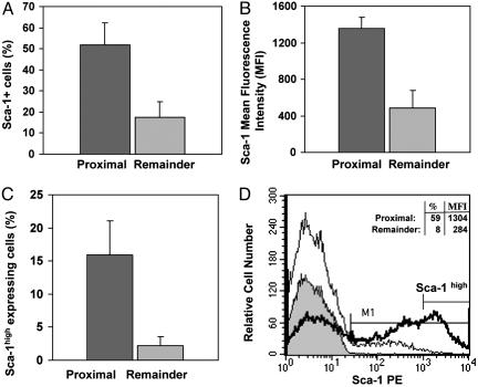
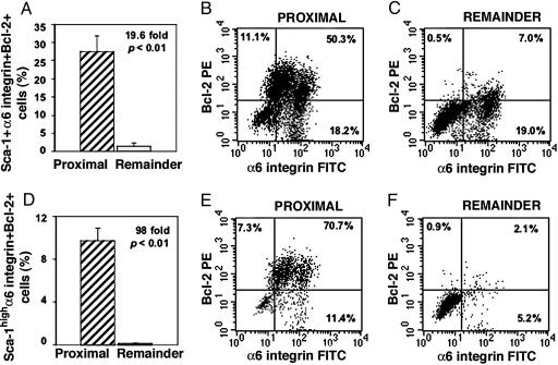
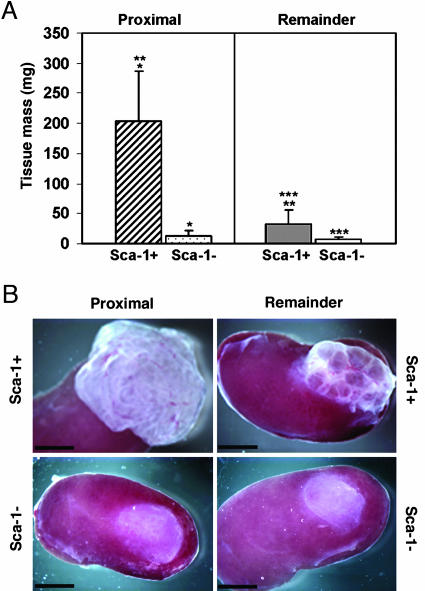
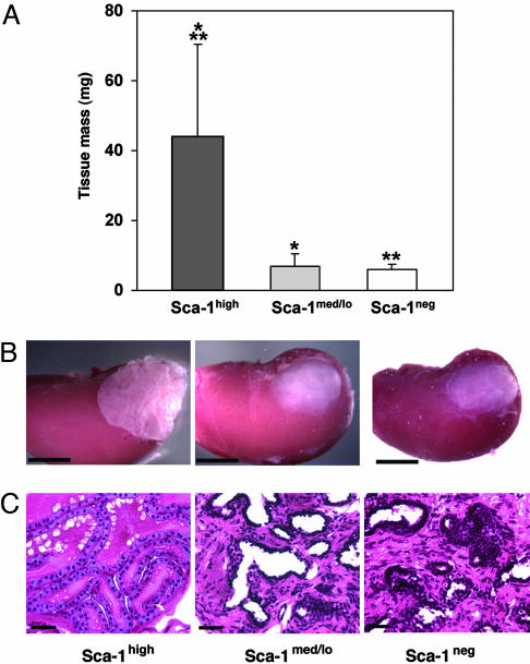
We previously showed that prostatic stem cells are concentrated in the proximal regions of prostatic ducts. We now report that these stem cells can be purified from isolated proximal duct regions by virtue of their high expression of the cell surface protein stem cell antigen 1 (Sca-1). In an in vivo prostate reconstitution assay, the purified Sca-1-expressing cell population isolated from the proximal region of ducts was more effective in generating prostatic tissue than a comparable population of Sca-1-depleted cells (203.0 +/- 83.1 mg vs. 11.9 +/- 9.2 mg) or a population of Sca-1-expressing cells isolated from the remaining regions of ducts (transit-amplifying cells) (31.9 +/- 24.1 mg). Almost all of the proliferative capacity of the proximal duct Sca-1-expressing cell population resides within the fraction of cells that express high levels of Sca-1 (top one-third), with the proximal region of prostatic ducts containing 7.2-fold more Sca-1(high) cells than the remaining regions. More than 60% of the high-expressing cells coexpress alpha6 integrin and the anti-apoptotic factor Bcl-2, markers that are also characteristic of stem cells of other origins. Further stratification of the phenotype of the stem cells may enable the development of rational therapies for treating prostate cancer and benign prostatic hyperplasia.


Figures

Fig. 1.
Fig. 1.
Sca-1 is highly expressed by cells in the proximal region of prostatic ducts. The expression of Sca-1 by cell digests from the proximal and remaining regions of prostatic ducts was determined. (A) The proximal region contained 2.9-fold more Sca-1-expressing cells than the remaining ductal regions (P < 0.0001). (B) Cells from the proximal region expressed 2.8-fold more molecules of Sca-1 per cell (higher MFI) than cells from the remaining regions (P < 0.01). (C) Cells with high levels of Sca-1 expression (cells with fluorescence intensities in the upper one-third) were 7.2-fold more prevalent in the proximal region than in the remaining regions (P < 0.00001). Results in A-C are means of at least three experiments. (D) A representative histogram (from one of five experiments) of Sca-1 expression by viable (7-aminoactinomycin D-negative) cells from the proximal region (thick line) and the remaining regions (thin line) of ducts shows the difference in Sca-1 expression between these two regions. The gray-filled histogram represents the appropriate IgG control. The marker M1 is placed so that <1% of control cells are positive. A second marker denotes Sca-1high cells.
Fig. 2.
Fig. 2.
The proximal region is considerably enriched in Sca-1-expressing cells that coexpress α6 integrin and Bcl-2. Three-color FACS analysis was performed to determine the incidence of Sca-1+ α6 integrin+ Bcl-2+ cells (A-C) and Sca-1high α6 integrin+ Bcl-2+ cells (D-F) in the proximal and remaining regions of ducts. (A) The proximal region contained 19.6-fold more Sca-1+ α6 integrin+ Bcl-2+ cells than the remaining regions (P < 0.01). (B and C) In these representative dot plots, 50.3% of proximal Sca-1+ cells coexpressed both α6 integrin and Bcl-2 (B), whereas 7.0% of cells from the remaining regions coexpressed these antigens (C). (D) Analysis of triple-labeled cells expressing high levels of Sca-1 showed that the proximal region contained 98-fold more Sca-1high α6 integrin+ Bcl-2+ cells than the remaining regions (P < 0.01). (E and F) For these dot plots, 70% of proximal Sca-1high cells coexpressed both α6 integrin and Bcl-2 (E), whereas 2% of cells from the remaining regions were Sca-1high α6 integrin+ Bcl-2+ (F). The results are the mean of three experiments.
Fig. 3.
Fig. 3.
Sca-1+ cells have greater in vivo proliferative capacity than Sca-1- cells. (A) The growth of Sca-1+ and Sca-1- cells (105 cells) that were isolated from either the proximal region or the remaining regions of ducts and transplanted under the renal capsule was measured after 8 weeks. Sca-1+ cells obtained from the proximal region formed 17.1-fold more prostatic tissue than Sca-1- cells (*, P < 0.001). Sca-1+ cells obtained from the remaining ductal regions had far less growth potential than Sca-1+ proximal cells (**, P < 0.001). Sca-1- cells from the remaining regions showed less growth than Sca-1+ cells from this region (***, P < 0.001). The results are the means of two experiments, using the data obtained from the inoculation of a total of 7, 11, 14, and 12 kidneys with Sca-1+ and Sca-1- cells from the proximal and remaining regions, respectively. (B) Prostate tissue under the renal capsule initiated with 105 Sca-1+ or Sca-1- cells from either the proximal region or the remaining regions. (Scale bars: 3 mm.)
Fig. 4.
Fig. 4.
Sca-1high cells have greater in vivo proliferative capacity than cells that express lower levels of Sca-1. Cells were isolated from the proximal region and sorted by FACS into Sca-1high, Sca-1med/lo and Sca-1neg fractions according to the level of Sca-1 expression. The cell populations (3 × 10⁴) were transplanted under the renal capsule and the growth of prostatic tissue was measured after 10 weeks. (A) Sca-1high cells formed 6.3-fold more prostatic tissue than Sca-1med/lo cells (*, P < 0.001) and 7.5-fold more prostatic tissue than Sca-1neg cells (**, P = 0.001). These results are the means of two experiments, using the data obtained from the inoculation of a total of 10, 9, and 10 kidneys with cell populations containing Sca-1high, Sca-1med/lo and Sca-1neg cells, respectively. (B) Prostate tissue initiated with 3 × 10⁴ Sca-1high, Sca-1med/lo, or Sca-1neg cells isolated by FACS from the proximal region of ducts. (Scale bars: 3 mm.) (C) Paraffin sections stained with hematoxylin and eosin showing the morphology of prostatic tissue arising from Sca-1high, Sca-1med/lo or Sca-1neg cells. The prostatic tissue obtained from Sca-1high cells had normal prostatic histology comprising basal and luminal cells lining prostatic ducts. The lumens of the ducts were filled with secretory material. The tissue arising from Sca-1med/lo and Sca-1neg cells contained increased stroma with less of an epithelial component, and little secretory material was noted within the ducts. (Scale bars: 40 μm.)

References

    1. Al-Hajj, M., Wicha, M. S., Benito-Hernandez, A., Morrison, S. J. & Clarke, M. F. (2003) Proc. Natl. Acad. Sci. USA 100, 3983-3988. - PMC - PubMed
    1. Al-Hajj, M. & Clarke, M. F. (2004) Oncogene 23, 7274-7282. - PubMed
    1. Lapidot, T., Sirard, C., Vormoor, J., Murdoch, B., Hoang, T., Caceres-Cortes, J., Minden, M., Paterson, B., Caligiuri, M. A. & Dick, J. E. (1994) Nature 367, 645-648. - PubMed
    1. Pardal, R., Clarke, M. F. & Morrison, S. J. (2003) Nat. Rev. Cancer 3, 895-902. - PubMed
    1. Reya, T., Morrison, S. J., Clarke, M. F. & Weissman, I. L. (2001) Nature 414, 105-111. - PubMed

Publication types