. 2005 Jun;3(6):e192.
doi: 10.1371/journal.pbio.0030192. Epub 2005 May 24.

Intravital observation of Plasmodium berghei sporozoite infection of the liver

Affiliations

Intravital observation of Plasmodium berghei sporozoite infection of the liver

Ute Frevert et al. PLoS Biol. 2005 Jun.

Abstract

Plasmodium sporozoite invasion of liver cells has been an extremely elusive event to study. In the prevailing model, sporozoites enter the liver by passing through Kupffer cells, but this model was based solely on incidental observations in fixed specimens and on biochemical and physiological data. To obtain direct information on the dynamics of sporozoite infection of the liver, we infected live mice with red or green fluorescent Plasmodium berghei sporozoites and monitored their behavior using intravital microscopy. Digital recordings show that sporozoites entering a liver lobule abruptly adhere to the sinusoidal cell layer, suggesting a high-affinity interaction. They glide along the sinusoid, with or against the bloodstream, to a Kupffer cell, and, by slowly pushing through a constriction, traverse across the space of Disse. Once inside the liver parenchyma, sporozoites move rapidly for many minutes, traversing several hepatocytes, until ultimately settling within a final one. Migration damage to hepatocytes was confirmed in liver sections, revealing clusters of necrotic hepatocytes adjacent to structurally intact, sporozoite-infected hepatocytes, and by elevated serum alanine aminotransferase activity. In summary, malaria sporozoites bind tightly to the sinusoidal cell layer, cross Kupffer cells, and leave behind a trail of dead hepatocytes when migrating to their final destination in the liver.


Figures

Figure 1
Figure 1. Architecture of the Liver Sinusoid
Liver sinusoids (S) are lined by fenestrated endothelia (EC) and interspersed Kupffer cells (KC), the resident macrophages of the liver. Stellate cells (SC), the major producers of liver ECM, are located inside the narrow space of Disse (D), which is formed by the sinusoidal cell layer and cords of hepatocytes (H).
Figure 2
Figure 2. Distribution of GFP-Expressing Endothelia and Kupffer Cells in the Liver
(A) Confocal microscopy demonstrates the GFP distribution in sinusoidal endothelia from a Tie2-GFP mouse. GFP is most prominent in the perinuclear region (arrowheads) of endothelia located in the periphery of the liver lobule. (B) A still image from an intravital movie shows GFP-expressing endothelia lining the sinusoids of a Tie2-GFP mouse. Kupffer cells can be identified by their orange autofluorescent lysosomes (arrowheads). (C) Star-shaped Kupffer cells (arrowheads) are located in sinusoids of a lys-EGFP-ki mouse liver. (D) Round blood granulocytes (arrows), traveling with the bloodstream or crawling along the sinusoidal cell layer, exhibit a stronger GFP signal than Kupffer cells (still image extracted from an intravital movie). Note the orange autofluorescence of the Kupffer cell lysosomes (arrowheads). Bar = 10 μm. See Videos S1 and S2.
Figure 3
Figure 3. Sporozoite Gliding along the Sinusoidal Endothelium
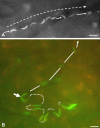
(A) A P. berghei sporozoite expressing fluorescent RedStar protein glides with and against the bloodstream inside a liver sinusoid of a Tie2-GFP mouse. The arrow indicates the overall movement of the parasite. (B) The projection through the same area of the liver visualizes the outline of the highly branched sinusoids. The direction of the blood flow is indicated by the dashed arrows. Bar = 10 μm. See Videos S3 and S4.
Figure 4
Figure 4. Sporozoite Passage into the Liver Parenchyma
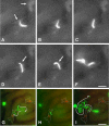
(A–E) show individual frames extracted from an intravital movie; (F) is a projection visualizing the transmigration path of the GFP P. berghei sporozoite in a Tie2-GFP mouse liver. (G–I) show projections from an intravital movie demonstrating the path of the parasite; its overall direction is indicated by arrows (dotted lines). (A) After gliding along a sinusoid, a sporozoite has encountered a Kupffer cell, which it faces with its apical cell pole. (B and C) Following a pause, the parasite slowly enters the Kupffer cell. (D and E) Sporozoite passage into the liver parenchyma occurs at a slow speed and involves the formation of a constriction in the parasite (arrow). (F) Once inside the liver tissue, the sporozoite increases its speed and transmigrates through several hepatocytes. (G) A projection from an intravital movie shows the path of a GFP P. berghei sporozoite gliding against the bloodstream along a sinusoid in a lys-EGFP-ki mouse liver. Eventually, the parasite encounters a Kupffer cell. (H) The sporozoite stops, facing the phagocyte with its apical cell pole. The outline of the two Kupffer cells in the image is indicated by dotted lines. (I) After slowly passing through the Kupffer cell, the sporozoite enters the liver parenchyma and migrates through several hepatocytes. Bars = 10 μm. See Videos S5 and S6.
Figure 5
Figure 5. P. berghei Sporozoite Transmigration Is Independent of the Species of the Infected Host
Projections of typical GFP P. berghei sporozoite paths show that the parasites transmigrate in a similar fashion through many hepatocytes in mouse ([A] Tie2-GFP mouse; [B] lys-EGFP-ki mouse) and also in rat livers (C and D). Bars = 10 μm. See Videos S7 and S8.
Figure 6
Figure 6. Unsuccessful Attempts of Liver Infection
(A) The composite image of ten selected frames from an intravital movie shows a paralyzed or dead P. berghei sporozoite that is eventually dislodged and flushed out of the liver lobule (short arrows). The parasite maintains a fixed crescent shape, fails to cling to the sinusoidal cell layer, and makes no attempt to glide against the bloodstream. The long arrow (dotted line) indicates the direction of the blood flow. (B) Projection composed of 14 selected frames extracted from an intravital movie showing a GFP P. berghei sporozoite that initially transmigrates in the liver parenchyma (dotted lines), but then reenters a sinusoid (arrow) and is swept away with the bloodstream (solid lines). Bars = 10 μm. See Videos S9 and S10.
Figure 7
Figure 7. Transmigrating Plasmodium Sporozoites Leave Behind a Trail of Dead Hepatocytes
(A) Three hours after infection with 5 × 10⁶ P. berghei salivary gland sporozoites, a mouse liver contains small clusters of necrotic hepatocytes that have been infiltrated by inflammatory cells (arrows). (B) Four hours after intravenous infection with 5 × 10⁶ P. yoelii sporozoites, a mouse liver contains individual or small clusters of necrotic hepatocytes (arrows). (C) Six hours after infection, the signs of hepatocytic damage appear more severe in another mouse liver (arrow). (D) Forty hours after inoculation of 2 × 10⁶ P. yoelii sporozoites, small infiltrates of inflammatory cells (arrows) block the lumina of some sinusoids, while maturing EEFs are free of any inflammatory reaction. (E) Fifty hours after infection with 2 × 10⁶ P. yoelii sporozoites, the size of the inflammatory infiltrates (arrows) has increased. Stains used: (A) paraffin section stained with H&E, (B and C) frozen sections stained with Evans blue, (D and E) semithin Epon sections stained with Toluidine blue. Bars = 20 μm.
Figure 8
Figure 8. Sporozoites, Surrounded by a Parasitophorous Vacuole Membrane, Can Be Found in Intact Hepatocytes
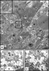
(A) Six hours after infection by bite of 50 P. yoelii–infected mosquitoes, electron microscopic examination of a mouse liver shows a sporozoite inside an intact hepatocyte. Note that the parasite is enclosed in a parasitophorous vacuole (insert). The neighboring hepatocyte shows signs of cytoplasmic swelling. (B and C) P. yoelii–infected mouse livers contain hepatocytes that exhibit various degrees of necrosis, ranging from hydropic swelling to near-complete disintegration of the cell, adjacent to parenchymal cells with a normal ultrastructure (arrows). The normal hepatocytes did not contain a sporozoite in the plane of the section. L, lipid droplet; M, mitochondrium; N, nucleus; S, sinusoid. Bars = 1 μm.
Figure 9
Figure 9. Sporozoite Transmigration Causes Histopathological Changes in the Liver
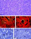
Mouse livers were removed 2 d (A–D) or 7 d (E–H) after daily infection with P. yoelii by bite of 150 mosquitoes and stained with H&E (A and E) or Masson's trichrome (B and F). Other sections were subjected to immunohistochemistry using mAb PC10 against proliferating cell nuclear antigen (C and G) or mAb HHF35 against smooth muscle actin (D and H). In contrast to the livers fixed after 2 d of infection, in which only a few cells reacted with mAb PC10 and mAb HHF35 (arrows in C and D), livers examined after 7 d of infection showed (E) increased numbers of nonparenchymal cells lining the sinusoids (arrow), (F) a focal deposition of collagen (blue) in some spaces of Disse, (G) large numbers of proliferating nonparenchymal cells and hepatocytes (brown, arrows), and (H) a focal increase in the concentration of smooth muscle actin (brown, arrow).
Figure 10
Figure 10. Sporozoite Infection Increases the Serum ALT Activity
(A) Three mice were inoculated with salivary gland extract from 100 uninfected mosquitoes each (labeled “0” on the x-axis). Another three mice were infected by intravenous inoculation into the tail vein of 0.7 × 10⁶, 1.2 × 10⁶, or 1.8 × 10⁶ purified P. yoelii sporozoites (indicated as “0.7,” “1.2,” and “1.8” on the x-axis). The ALT activity in the serum was determined before and after infection at the indicated time points. In comparison to the control serum drawn before infection, the ALT levels increased significantly and depended on the number of inoculated sporozoites in all mice during the observation period of 52 h. Uninfected salivary gland extract had only a temporary effect on the serum ALT level (9 h). The indicated values represent the average ± standard deviation of triplicate measurements. *, p < 0.005 in relation to the corresponding control sera. (B) No change in the serum ALT activity was detectable when three mice were infected with P. berghei by bite of 150 mosquitoes. The indicated values represent the average ± standard deviation of duplicate measurements. For ready comparison, the data were normalized for each animal prior to statistical analysis.
Figure 11
Figure 11. Model of Plasmodium Sporozoite Infection of the Mammalian Liver
The dual blood supply of the liver, consisting of branches of the portal vein and the hepatic artery, merges upon entry into the liver lobule at the portal field. The blood flows along the sinusoid and exits at the central vein. First sporozoites enter the liver lobule either via the portal vein or the hepatic artery, and then are abruptly arrested by binding to the sinusoidal cell layer. The initial binding is presumably mediated by stellate-cell-derived ECM proteoglycans that protrude from the space of Disse across the endothelial sieve plates into the sinusoidal lumen. After a pause, the parasites begin to glide along the sinusoid, frequently moving against the bloodstream, until they then encounter a Kupffer cell, on the surface of which they recognize selected chondroitin and heparan sulfate proteoglycans. Sporozoites position themselves with their apical cell pole facing the phagocyte. After a considerable pause, they slowly pass through the Kupffer cell and cross the space of Disse beyond it, exhibiting a clearly visible constriction. Once inside the liver parenchyma, the parasites increase their velocity and migrate for many minutes through several hepatocytes, before they eventually settle down in a final one for EEF development. Sporozoite transmigration results in a trail of necrotic hepatocytes, whose remains are subsequently removed by infiltrating inflammatory cells.

References

    1. Vanderberg JP, Frevert U. Intravital microscopy demonstrating antibody-mediated immobilization of Plasmodium berghei sporozoites injected into skin by mosquitoes. Int J Parasitol. 2004;34:991–996. - PubMed
    1. Cerami C, Frevert U, Sinnis P, Takacs B, Clavijo P, et al. The basolateral domain of the hepatocyte plasma membrane bears receptors for the circumsporozoite protein of Plasmodium falciparum sporozoites. Cell. 1992;70:1021–1033. - PubMed
    1. Ying P, Shakibaei M, Patankar MS, Clavijo P, Beavis RC, et al. The malaria circumsporozoite protein: Interaction of the conserved regions I and II-plus with heparin-like oligosaccharides in heparan sulfate. Exp Parasitol. 1997;85:168–182. - PubMed
    1. Pradel G, Garapaty S, Frevert U. Proteoglycans mediate malaria sporozoite targeting to the liver. Mol Microbiol. 2002;45:637–651. - PubMed
    1. Frevert U, Sinnis P, Cerami C, Shreffler W, Takacs B, et al. Malaria circumsporozoite protein binds to heparan sulfate proteoglycans associated with the surface membrane of hepatocytes. J Exp Med. 1993;177:1287–1298. - PMC - PubMed

Publication types

Associated data