. 2005 Sep 1;390(Pt 2):455-64.
doi: 10.1042/BJ20050113.

The signal peptide of the rat corticotropin-releasing factor receptor 1 promotes receptor expression but is not essential for establishing a functional receptor

Affiliations

The signal peptide of the rat corticotropin-releasing factor receptor 1 promotes receptor expression but is not essential for establishing a functional receptor

Martina Alken et al. Biochem J. .

Abstract

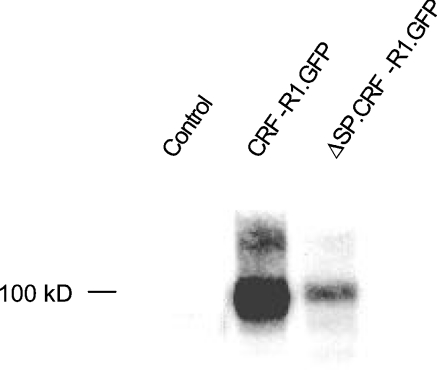
Approximately 5-10% of the GPCRs (G-protein-coupled receptors) contain N-terminal signal peptides that are cleaved off during receptor insertion into the ER (endoplasmic reticulum) membrane by the signal peptidases of the ER. The reason as to why only a subset of GPCRs requires these additional signal peptides is not known. We have recently shown that the signal peptide of the human ET(B)-R (endothelin B receptor) does not influence receptor expression but is necessary for the translocation of the receptor's N-tail across the ER membrane and thus for the establishment of a functional receptor [Köchl, Alken, Rutz, Krause, Oksche, Rosenthal and Schülein (2002) J. Biol. Chem. 277, 16131-16138]. In the present study, we show that the signal peptide of the rat CRF-R1 (corticotropin-releasing factor receptor 1) has a different function: a mutant of the CRF-R1 lacking the signal peptide was functional and displayed wild-type properties with respect to ligand binding and activation of adenylate cyclase. However, immunoblot analysis and confocal laser scanning microscopy revealed that the mutant receptor was expressed at 10-fold lower levels than the wild-type receptor. Northern-blot and in vitro transcription translation analyses precluded the possibility that the reduced receptor expression is due to decreased transcription or translation levels. Thus the signal peptide of the CRF-R1 promotes an early step of receptor biogenesis, such as targeting of the nascent chain to the ER membrane and/or the gating of the protein-conducting translocon of the ER membrane.


Figures

Figure 1
Figure 1. Amino acid sequence of the N-tail of CRF-R1 and schematic representation of the CRF-R1 constructs used in the present study
(A) Sequence of the N-tail of the CRF-R1 and its cleavable signal peptide (black). (B) Full-length receptor constructs. CRF-R1.GFP and ΔSP.CRF-R1.GFP represent the wild-type GFP-tagged CRF-R1 and its signal peptide mutant respectively. The signal peptide and TMs (numbered) are shown as black boxes. The N-tail is depicted as an open box. (C) Marker protein fusions. NT.GFP and ΔSP.NT.GFP are fusion proteins consisting of GFP and the N-tail with and without signal peptide respectively. For purification, an additional C-terminal His6-sequence (His) is added. Plasmids NT.AP and ΔSP.NT.AP represent the corresponding AP fusions. Plasmid CRF-R1.PrP encodes a fusion of the N-tail of CRF-R1 to the hamster prion marker protein. Plasmid ΔSP.CRF-R1.PrP encodes the corresponding signal peptide mutant.
Figure 2
Figure 2. CRF-R1 possesses a cleavable signal peptide
(A) Schematic representation of a method to detect cleavable signal peptides of membrane proteins. Putative signal peptides are fused to marker proteins (AP or GFP). In the presence of a signal peptide, the marker proteins are directed via the secretory pathway to the extracellular space. Secreted marker proteins are detectable by their autofluorescence (GFP) or by an enzyme activity assay (AP). Ri, ribosome. (B) Upper panel: GFP fluorescence assay. HEK-293 cells were transiently transfected with fusion proteins consisting of GFP and the N-tail of CRF-R1 with signal peptide (NT.GFP) and without signal peptide (ΔSP.NT.GFP). Untransfected HEK-293 cells were used as a control. The fusions proteins were purified from the supernatant and the GFP fluorescence signals were monitored. Results shown are the means±S.D. for three independent experiments each performed in triplicates. Lower panel: detection of the same purified fusion proteins by immunoblotting using a polyclonal rabbit anti-GFP antibody and AP-conjugated anti-rabbit IgG. (C) Upper panel: AP activity assay. HEK-293 cells were transiently transfected with fusion proteins consisting of AP and the N-tail of CRF-R1 with signal peptide (NT.AP) and without signal peptide (ΔSP.NT.AP). Untransfected HEK-293 cells were used as a control. The AP activity of each supernatant was quantified by an enzymatic assay. Results shown are the means±S.D. for three independent experiments each performed in triplicates. Lower panel: detection of the same fusion proteins by immunoblotting using a monoclonal mouse anti-His antibody and AP-conjugated anti-mouse IgG. The fusion proteins were purified before SDS/PAGE/immunoblotting.
Figure 3
Figure 3. Pharmacological properties of the GFP-tagged CRF-R1 and ETB-R and their respective signal peptide mutants
(A) Left panel: [125I]Tyr0–sauvagine-binding profiles of intact transiently transfected HEK-293 cells expressing CRF-R1.GFP (wild-type CRF-R1) and the signal peptide mutant ΔSP.CRF-R1.GFP. Specific binding is shown. Data points represent the means±S.E.M. for three independent experiments each performed in triplicates. Unspecific binding contributed up to 10% of the total binding. The calculated KD and Bmax values are indicated. Right panel: adenylate cyclase activity assay with crude membranes of transiently transfected HEK-293 cells expressing the same constructs as in the left-hand panel. Data points represent the means±S.E.M. for three independent experiments each performed in triplicates. The calculated EC50 values are indicated. (B) Left panel: [125I]ET-1-binding profiles of intact transiently transfected HEK-293 cells expressing ETB.GFP (wild-type ETB-R) and its signal peptide mutant ETBΔ26.GFP. Specific binding is shown. Data points represent the means±S.E.M. for three independent experiments each performed in triplicates. Unspecific binding contributed up to 15% of the total binding. The calculated KD and Bmax values are indicated. Right panel: ET-1-mediated IP accumulation in intact transiently transfected HEK-293 cells expressing the same constructs as in the left-hand panel. Data points represent the means±S.E.M. derived from a single experiment performed in triplicate. The dose–response curve is representative of two independent experiments. The calculated EC50 values are indicated.
Figure 4
Figure 4. Cell-surface biotinylation assay with transiently transfected HEK-293 cells expressing CRF-R1.GFP and its signal peptide mutant ΔSP.CRF-R1.GFP
Plasma-membrane proteins of intact cells were labelled with biotin. Biotinylated proteins were isolated with NeutrAvidin, and labelled receptors were detected by immunoblotting, using a polyclonal rabbit anti-GFP antiserum and peroxidase-conjugated anti-rabbit IgG. Untransfected HEK-293 cells were used as a control. The immunoblot is representative of three independent experiments.
Figure 5
Figure 5. Confocal laser scanning microscopy of transiently transfected HEK-293 cells expressing CRF-R1.GFP and its signal peptide mutant ΔSP.CRF-R1.GFP
(A) Localization of the GFP fluorescence signals. Shown are horizontal (xy) scans. GFP signals are shown in green (left panels) and Trypan Blue signals of the cell surface of the same cells in red (central panels). GFP and Trypan Blue fluorescence signals were computer-overlayed (right panels; overlap is indicated by yellow). The scans show representative cells. Scale bar, 10 μm. Similar data were obtained in three independent experiments. (B) Quantification of the plasma-membrane GFP fluorescence signals of the cells. The GFP signals were analysed by horizontal scans (left panels). The GFP signal intensities were measured using an 8 bit grey scale (ranging from 0 to 250) and transformed into a colour scale (central panel; see the Figure for colour scale). The coloured signal intensities are also shown using a three-dimensional plot of the cells (right panels). (C) Statistical analysis. Results shown are the means±S.D. for the plasma-membrane GFP fluorescence intensities of cells (n=30). Fluorescence intensities of the individual cells were recorded as shown in (B).
Figure 6
Figure 6. Immunoprecipitation of GFP-tagged CRF-R1 and ETB-R constructs and their respective signal peptide mutants
(A) CRF-R1 constructs. Crude membranes of transiently transfected HEK-293 cells expressing CRF-R1.GFP and ΔSP.CRF-R1.GFP were isolated and receptors were immunoprecipitated using a polyclonal rabbit anti-GFP antiserum. Precipitated receptors were detected by immunoblotting using a monoclonal mouse anti-GFP antibody and peroxidase-conjugated anti-mouse IgG. Membranes were either treated with PNGase F to remove N-glycosylations (+) or left untreated (−). The immunoblot is representative of three independent experiments. #, complex-glycosylated receptors; >, high-mannose forms; *, non-glycosylated receptors. (B) ETB-R constructs. Crude membranes of transiently transfected HEK-293 cells expressing ETB.134.GFP and ETB134Δ26.GFP were isolated. Receptor fragments were immunoprecipitated and detected as described above. Membranes were either treated with PNGase F to remove N-glycosylations (+) or left untreated (−). The immunoblot is representative of two independent experiments.
Figure 7
Figure 7. Northern-blot analysis and translation assay
(A) Northern-blot analysis of CRF-R1.GFP and the signal peptide mutant ΔSP.CRF-R1.GFP. Total RNA was isolated from transiently transfected HEK-293 cells and Northern-blot analysis (20 μg of RNA/lane) was performed with 32P-labelled CRF-R1 cDNA. Blots were stripped and reprobed with a 32P-labelled cDNA fragment specific for actin. Untransfected cells were used as a control. The Northern blot is representative of three independent experiments. (B) In vitro transcription/translation assay of the N-tail of CRF1-R1 with and without signal peptide fused to the hamster PrP as a marker (constructs CRF-R1.PrP and ΔSP.CRF-R1.PrP respectively). [35S]Met was incorporated into the proteins using SP6 RNA polymerase and rabbit reticulocyte lysate. Labelled proteins were detected by audioradiography. The experiment is representative of two independent experiments.

Similar articles

Cited by

References

    1. Wallin E., von Heijne G. Properties of N-terminal tails in G protein-coupled receptors: a statistical study. Protein Eng. 1995;8:693–698. - PubMed
    1. Martoglio B., Dobberstein B. Signal peptide fragments of preprolactin and HIV-1 p-gp160 interact with calmodulin. Trends Cell Biol. 1998;8:410–415. - PMC - PubMed
    1. Köchl R., Alken M., Rutz C., Krause G., Oksche A., Rosenthal W., Schülein R. The signal peptide of the G protein-coupled human endothelin B receptor is necessary for translocation of the N terminal tail across the endoplasmic reticulum membrane. J. Biol. Chem. 2002;277:16131–16138. - PubMed
    1. Rapoport T. A., Jungnickel B., Kutay U. Protein transport across the eukaryotic endoplasmic reticulum and bacterial inner membranes. Annu. Rev. Biochem. 1996;65:271–303. - PubMed
    1. Hegde R. S., Lingappa V. R. Regulation of protein biogenesis at the endoplasmic reticulum membrane. Trends Cell Biol. 1999;9:132–137. - PubMed

Publication types

Substances