. 2005 Jun;132(12):2733-42.
doi: 10.1242/dev.01865. Epub 2005 May 18.

Xema, a foxi-class gene expressed in the gastrula stage Xenopus ectoderm, is required for the suppression of mesendoderm

Affiliations

Xema, a foxi-class gene expressed in the gastrula stage Xenopus ectoderm, is required for the suppression of mesendoderm

Crystal Suri et al. Development. 2005 Jun.

Abstract

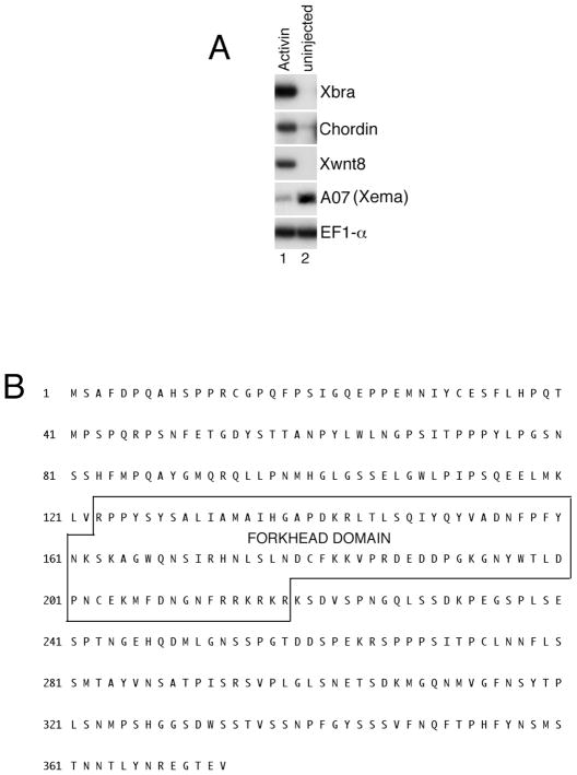
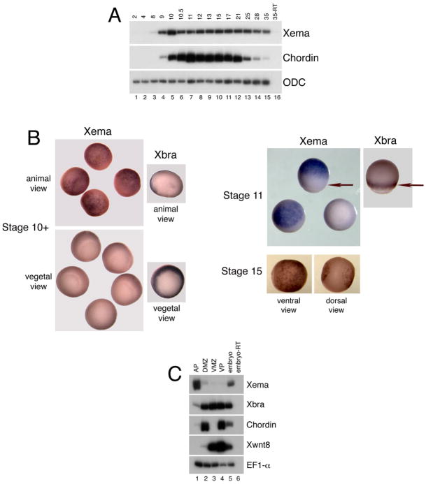
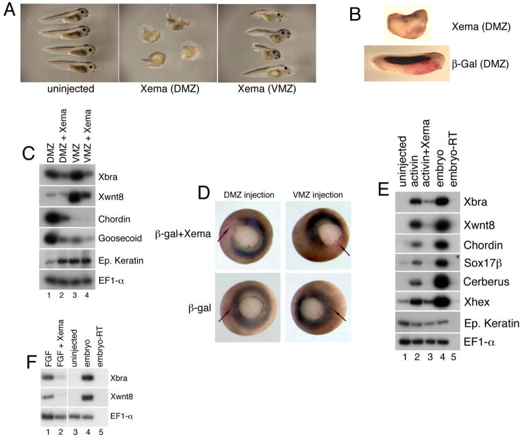
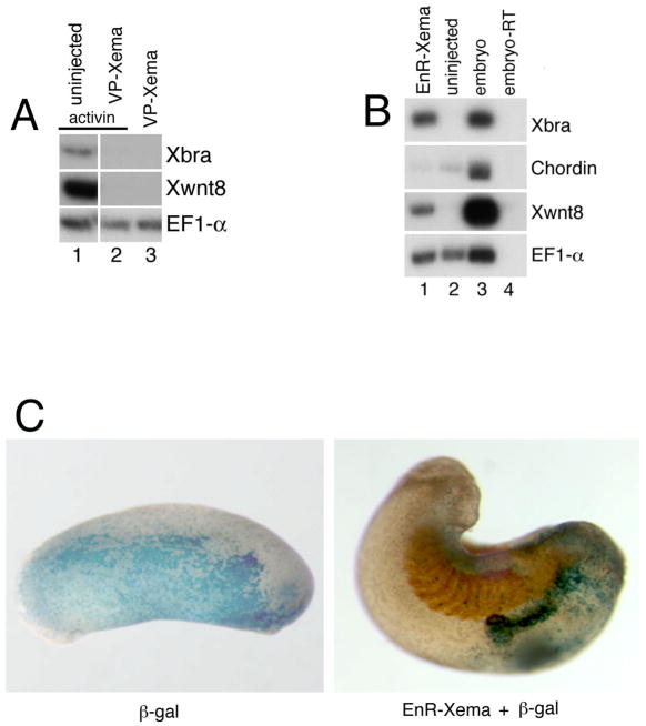
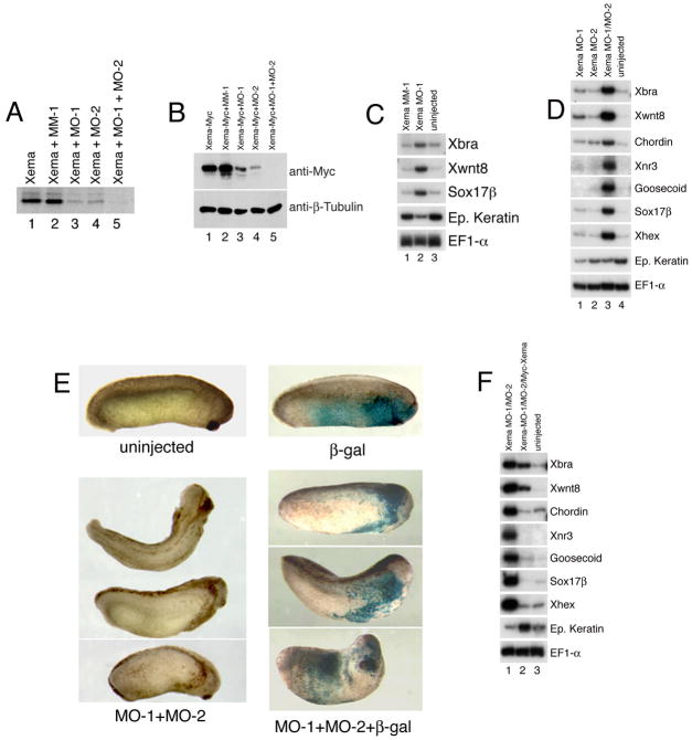
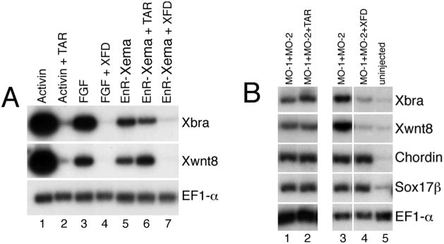
The molecular basis of vertebrate germ layer formation has been the focus of intense scrutiny for decades, and the inductive interactions underlying this process are well defined. Only recently, however, have studies demonstrated that the regulated inhibition of ectopic germ layer formation is also crucial for patterning the early vertebrate embryo. We report here the characterization of Xema (Xenopus Ectodermally-expressed Mesendoderm Antagonist), a novel member of the Foxi-subclass of winged-helix transcription factors that is involved in the suppression of ectopic germ layer formation in the frog, Xenopus laevis. Xema transcripts are restricted to the animal pole ectoderm during early Xenopus development. Ectopic expression of Xema RNA inhibits mesoderm induction, both by growth factors and in the marginal zone, in vivo. Conversely, introduction of antisense morpholino oligonucleotides directed against the Xema transcript stimulates the expression of a broad range of mesodermal and endodermal marker genes in the animal pole. Our studies demonstrate that Xema is both necessary and sufficient for the inhibition of ectopic mesendoderm in the cells of the presumptive ectoderm, and support a model in which Fox proteins function in part to restrict inappropriate germ layer development throughout the vertebrate embryo.


Figures

Fig. 1
Fig. 1
Identification of the Xenopus Foxi-class gene Xema. (A) Activin inhibits A07 (Xema) expression. RT-PCR analysis of animal cap explants dissected at late blastula stages and cultured until midgastrula stages. Activin (0.5 ng/ml) was added to stage 9 animal caps, as listed. (B) Putative Xema amino acid sequence. The forkhead box (Fox) DNA-binding domain is indicated.
Fig. 2
Fig. 2
Expression of Xema during early development. (A) RT-PCR analysis of Xema temporal expression. The ‘-RT’ lane contains all reagents except reverse transcriptase and was used as a negative control. Ornithine decarboxylase (ODC) is used as a loading control (Bassez et al., 1990). Chordin expression is initiated zygotically and is used as a positive control. (B) Whole-mount in situ hybridization of early gastrula (Stage 10+), midgastrula (Stage 11), and early neurula (Stage 15) stage embryos, using antisense Xema and Xbrachyury (Xbra) probes. (B) Xema expression is seen as a blue stain throughout the animal pole of gastrula stage albino embryos; Xema expression is excluded from the marginal zone (denoted by arrows at stage 11) and vegetal pole. Expression of the panmesodermal marker Xbra is found only in the marginal zone of gastrula stage embryos and is used as a control. Xema is expressed in the vental ectoderm (epidermis) of early neurula stage embryos (ventral view); expression is excluded from the neural plate (dorsal view). (C) RT-PCR analysis of Xema expression in early gastrula stage explants. EF1-α is used as a loading control (Krieg et al., 1989). Xbra is a panmesodermal marker at this stage (Smith et al., 1991); chordin is a dorsal endomesodermal marker (Sasai et al., 1994); Xwnt8 is a ventrolateral marker (Christian et al., 1991; Smith and Harland, 1991).
Fig. 3
Fig. 3
Ectopic Xema inhibits mesendoderm induction. (A) Xema misexpression disrupts embryonic development. Lateral views of stage 35 embryos, anterior is to right. (B) Whole-mount immunohistochemistry of tailbud stage embryos using the somite-specific antibody 12/101; lateral views, anterior is to right. Embryos were stained with Red Gal as a substrate prior to immunohistochemistry. (C) RT-PCR analysis of dorsal (DMZ) and ventral (VMZ) marginal zone explants from embryos injected with Xema RNA, harvested immediately after dissection at early gastrula stages. (D) Whole-mount in situ hybridization of midgastrula stage embryos, using an antisense Xbrachyury probe; vegetal pole views. Embryos were stained with Red Gal as a substrate prior to in situ hybridization. The embryos shown at the top of the figure were co-injected with β-galactosidase RNA and Xema RNA in two dorsal or ventral blastomeres, at the 4-cell stage, as listed; note the presence of β-galactosidase activity (red) in the gap in Xbrachyury expression (blue). The embryos shown at the bottom of the figure were injected with β-galactosidase RNA in the same regions, as indicated; note the overlap between Xbrachyury expression and β-galactosidase activity. (E) Inhibition of Activin-mediated mesendoderm induction by Xema. RT-PCR analysis of animal cap explants dissected at late blastula stages and cultured until midgastrula stages. Xema RNA was injected into the animal pole region of both blastomeres at the 2-cell stage. Activin (0.5 ng/ml) was added to stage 9 animal caps, as listed. (F) Inhibition of FGF-mediated mesoderm induction by Xema. bFGF (10 ng/ml) was added to stage 9 animal caps, as listed. For all experiments in this Figure, 1 ng of Xema RNA, and/or 100 pg β-Galactosidase RNA was injected, as listed.
Fig 4
Fig 4
Activity of Xema activator and repressor constructs. (A) VP-Xema-mediated inhibition of mesoderm formation. RT-PCR analysis of animal caps dissected at late blastula stages and cultured until midgastrula stages. 1 ng of VP-Xema RNA was injected at early cleavage stages, as listed. Activin (0.5 ng/ml) was added to stage 9 animal caps, as listed. (B) Expression of EnR-Xema induces mesoderm. RT-PCR analysis of animal caps dissected at late blastula stages and cultured until midgastrula stages. 1 ng of EnR-Xema RNA was injected at early cleavage stages, as listed. (C) Expression of EnR-Xema induces ectopic structures. Whole-mount immunohistochemistry of tailbud stage embryos using the somite-specific antibody 12/101; lateral views, anterior is to right. Embryos were stained with X-Gal as a substrate prior to immunohistochemistry. The embryo on the right was co-injected with 100pg β-Galactosidase and 1ng Xema RNA in the animal pole at the 2-cell stage; note the absence of somite staining (red) in the secondary structure containing the β-galactosidase activity (blue). The embryo on the left was injected with 100pg β-Galactosidase RNA in the animal pole at the 2-cell stage and was not probed with the somite-specific antibody.
Fig 5
Fig 5
Knockdown of Xema protein induces mesoderm formation. (A) Xema morpholinos block the translation of Xema RNA in vitro. Morpholino oligos (500 ng each) and Xema RNA (1 ng) were simultaneously added to a rabbit reticulocyte lysate mix (Promega), in the presence of [35S]Methionine. (B) Effect of Xema morpholinos on exogenous Xema protein levels. Western blot analysis of whole cell lysates extracted from embryos injected with 1ng Xema-Myc RNA. 10 ng MO-1, 10 ng MM-1, and/or 20 ng MO-2 were injected, as listed. (C) Mesoderm and endoderm are induced by injection of Xema MO-1 (10 ng) but not by Xema MM-1 (10 ng). RT-PCR analysis of animal cap explants from embryos injected with Xema morpholinos in the animal pole at early cleavage stages, dissected at blastula stages and harvested during midgastrula stages. (D) Xema MO-1 (10 ng) and MO-2 (20 ng) potently synergize to induce mesendoderm. (E) Co-expression of Xema MO-1 and MO-2 disrupts normal development. The lower left panels show embryos co-injected with 5 ng of MO-1 and 10 ng of MO-2 in the animal pole of early cleavage stage embryos. The top left embryo is uninjected. Second embryo from top is a dorsal view; all other views are lateral; anterior is to right. Note anterior pigmented ectopic lateral structures. The lower right panels show embryos injected with 5 ng of MO-1, 10 ng MO-2, and 166 pg of β-galactosidase RNA in the animal pole of early cleavage embryos. The top embryo is injected with 166 pg of β-galactosidase RNA only. Note the presence of β-galactosidase staining (blue), primarily in the ectopic structures. All views are lateral; anterior is to right. (F) Rescue of Xema morpholino-mediated mesendoderm formation by injection of 1 ng Myc-Xema RNA. 10 ng MO-1 and 20 ng MO-2 were injected, as listed.
Fig 6
Fig 6
FGF signaling is required for mesoderm induction by either EnR-Xema or Xema morpholinos. (A) EnR-Xema activity is inhibited by co-expression of a truncated FGF receptor (XFD), but is not inhibited by expression of a truncated activin receptor (TAR). RT-PCR analysis of animal cap explants dissected at blastula stages and harvested during midgastrula stages. 1 ng of EnR-Xema RNA, alone or with either 4 ng XFD or 4 ng TAR, was injected at early cleavage stages, as listed. Activin (0.5 ng/mL) or FGF (10 ng/mL) were added to stage 9 animal caps, as listed. (B) XFD inhibits Xema morpholino-mediated Xbra and Xwnt8 expression; TAR did not inhibit Xbra and Xwnt8 expression in 9/12 independent trials. Neither TAR nor XFD inhibit dorsal endomesodermal (chordin) or endodermal (sox17β) marker induction by Xema morpholino injection. 4 ng TAR, 4 ng XFD, 10 ng MO-1, and 20 ng MO-2 were injected at early cleavage stages, as listed.

Similar articles

Cited by

References

    1. Amaya E, Musci TJ, Kirschner MW. Expression of a dominant negative mutant of the FGF receptor disrupts mesoderm formation in Xenopus embryos. Cell. 1991;66:257–70. - PubMed
    1. Ang SL, Rossant J. HNF-3 beta is essential for node and notochord formation in mouse development. Cell. 1994;78:561–74. - PubMed
    1. Bassez T, Paris J, Omilli F, Dorel C, Osborne HB. Post-transcriptional regulation of ornithine decarboxylase in Xenopus laevis oocytes. Development. 1990;110:955–62. - PubMed
    1. Beddington RS, Robertson EJ. Axis development and early asymmetry in mammals. Cell. 1999;96:195–209. - PubMed
    1. Bouwmeester T, Kim S, Sasai Y, Lu B, De Robertis EM. Cerberus is a head-inducing secreted factor expressed in the anterior endoderm of Spemann’s organizer. Nature. 1996;382:595–601. - PubMed

Publication types

MeSH terms

Associated data

LinkOut - more resources