. 2005 Jun 1;19(11):1294-305.
doi: 10.1101/gad.1304105. Epub 2005 May 18.

Proapoptotic Bak is sequestered by Mcl-1 and Bcl-xL, but not Bcl-2, until displaced by BH3-only proteins

Affiliations

Proapoptotic Bak is sequestered by Mcl-1 and Bcl-xL, but not Bcl-2, until displaced by BH3-only proteins

Simon N Willis et al. Genes Dev. .

Abstract

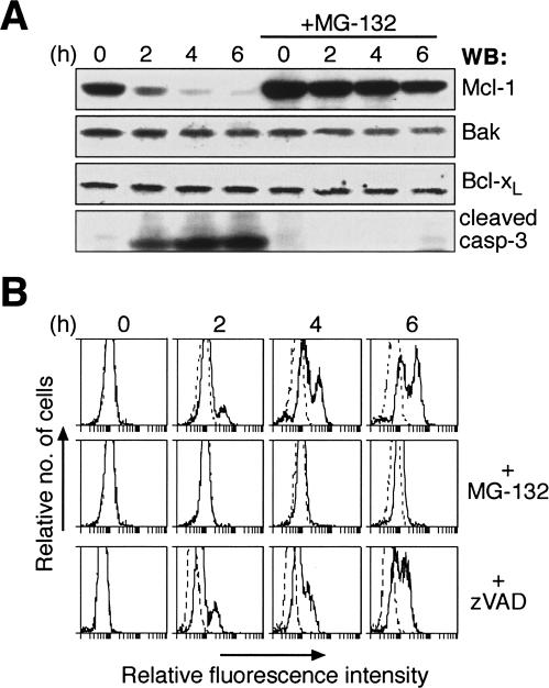
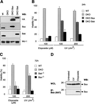
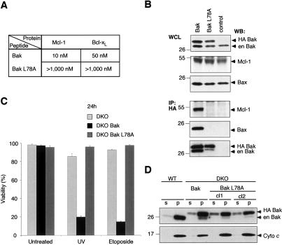
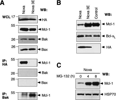
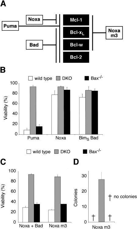
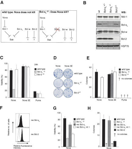
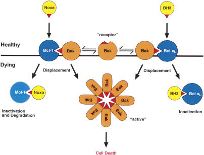
Commitment of cells to apoptosis is governed largely by the interaction between members of the Bcl-2 protein family. Its three subfamilies have distinct roles: The BH3-only proteins trigger apoptosis by binding via their BH3 domain to prosurvival relatives, while the proapoptotic Bax and Bak have an essential downstream role involving permeabilization of organellar membranes and induction of caspase activation. We have investigated the regulation of Bak and find that, in healthy cells, Bak associates with Mcl-1 and Bcl-x(L) but surprisingly not Bcl-2, Bcl-w, or A1. These interactions require the Bak BH3 domain, which is also necessary for Bak dimerization and killing activity. When cytotoxic signals activate BH3-only proteins that can engage both Mcl-1 and Bcl-x(L) (such as Noxa plus Bad), Bak is displaced and induces cell death. Accordingly, the BH3-only protein Noxa could bind to Mcl-1, displace Bak, and promote Mcl-1 degradation, but Bak-mediated cell death also required neutralization of Bcl-x(L) by other BH3-only proteins. The results indicate that Bak is held in check solely by Mcl-1 and Bcl-x(L) and induces apoptosis only if freed from both. The finding that different prosurvival proteins have selective roles has notable implications for the design of anti-cancer drugs that target the Bcl-2 family.


Figures

Figure 1.
Figure 1.
UV irradiation promotes Mcl-1 degradation to trigger Bak activation. (A) Proteasome inhibition prevents UV-induced Mcl-1 degradation. Lysates prepared from untreated or UV-irradiated (200 J/m2) HeLa cells were resolved by SDS-PAGE and the resulting blot probed with the indicated antibodies. (Right panels) The rapid Mcl-1 degradation and caspase-3 cleavage after UV was blocked in cells pretreated with the proteasome inhibitor MG-132. (B) Abrogation of UV-induced Bak activation by proteasome inhibition. Bak activation detected by flow cytometric analysis of untreated or UV-irradiated HeLa cells stained with an antibody (Ab-1) that specifically recognizes activated Bak (Griffiths et al. 1999). Some cells were pretreated with the proteasome inhibitor MG-132 (middle) or the broad-spectrum caspase inhibitor zVAD.fmk (bottom). Controls (dotted histograms) represent cells stained with the secondary antibody alone.
Figure 2.
Figure 2.
UV irradiation kills MEFs predominantly by a Bak-dependent, not Bax-dependent, mechanism. (A) Expression of Bax or Bak in MEFs. Immunoblot analysis of lysates prepared from immortalized wild-type MEFs, ones lacking Bax and Bak (DKO), or DKO subclones reconstituted with HA-tagged Bax (DKO Bax) or Bak (DKO Bak), using antibodies to HA (to specifically detect transgene expression), Bax, Bak, or Mcl-1. (B) Killing of immortalized MEFs by UV depends primarily on Bak, rather than Bax. Whereas exposure to 100 μM etoposide for 24 h caused comparable killing of Bax- or Bak-expressing MEFs (described in A), far more Bak-expressing than Bax-expressing cells died 24 h after exposure to UV (doses indicated). (C) UV-induced killing of primary MEFs is mainly mediated by Bak. Primary MEFs (derived independently from those used in A,B) were challenged with UV or etoposide. In B and C, cell viability was assessed by flow cytometric analyses after staining with propidium iodide (PI); the data represent mean ± SD from three independent experiments. (D) Unlike etoposide treatment, UV does not cause significant Bax activation in transformed MEFs. Lysates prepared from untreated Bak-/- MEFs, or 24 h after UV or etoposide treatment, were immunoblotted for total Bax (top) or for activated Bax (bottom) after immunoprecipitating with the conformation-specific antibody 6A7.
Figure 3.
Figure 3.
Bak is sequestered by Mcl-1 and Bcl-xL in healthy cells. (A) Tight binding of Bak BH3 to Mcl-1 and Bcl-xL. Using solution competition assays, the relative affinities (IC50 in nanomolar) of a BakBH3 peptide for prosurvival Bcl-2 proteins were determined (Chen et al. 2005). The results (plotted on an inverse log scale) are from representative experiments; the variation observed in multiple experiments was less than two-fold (using different chips or protein batches). (†) IC50 > 1000 nM. (B) Overexpressed Mcl-1 and Bcl-xL bind endogenous Bak. N-terminally Flag-tagged prosurvival proteins were overexpressed (top) in 293T cells and their capacity to bind endogenous Bak (middle) was tested by coimmunoprecipitation (bottom) using an anti-Flag affinity resin. (Control) Immunoprecipitation from untransfected cells; (en) endogenous; () an Mcl-1 break-down product; (★★) immunoglobulin light chain from the immunoprecipitating antibody. (C) Mcl-1, Bcl-xL, and Bak are present in the pellet fraction of healthy cells. HeLa cells, lysed in 0.025% digitonin, were fractionated into soluble (s) and pellet (p) fractions, and probed for the indicated proteins. Note that Bax, unlike Bak, is present mainly in the soluble fraction. (D) Endogenous Bcl-xL and Mcl-1 associate with endogenous Bak in healthy cells. HeLa cells (lysed in 0.025% digitonin) were fractionated into soluble (s) and pellet (p) fractions. The pellet fraction was solubilized in buffer containing Triton X-100, immunoprecipitated with anti-Mcl-1 (left), anti-Bcl-xL (right), or isotype-matched control antibodies, and examined for the presence of Bak (middle) or Bax (bottom).
Figure 4.
Figure 4.
Bak BH3 is required for interaction with Mcl-1 and Bcl-xL, and for proapoptotic function. (A) A point mutation within Bak BH3 abrogates interaction with Mcl-1 and Bcl-xL. Using solution competition assays, the relative affinities (IC50 in nanomolar) of Bak and mutant Bak L78A peptides for Mcl-1 and Bcl-xL were determined. (B) Bak L78A fails to heterodimerize with Mcl-1 or homodimerize. N-terminally HA-tagged wild-type Bak or mutant Bak L78A were transiently expressed in 293T cells (top) and tested for their ability to bind endogenous Mcl-1, Bax or Bak (bottom) by coimmunopreciptation using anti-HA affinity resin. (Control) Immunoprecipitation from untransfected cells; (en) endogenous. (C) L78A mutation inactivates Bak proapoptotic function. Viability was determined for Bax/Bak-deficient (DKO) MEFs or ones containing introduced Bak or Bak L78A, left untreated or 24 h after UV or etoposide treatment. Data represent mean ± SD from three independent experiments. (D) L78A mutant Bak, like wild-type Bak, localizes to the pellet fraction. Wild-type MEFs or Bax/Bak-deficient ones expressing wild-type Bak or mutant Bak L78A (two independent clones) were fractionated (in digitonin-containing buffer) into soluble (s) and pellet (p) fractions, and probed for Bak (top) or cytochrome c (bottom).
Figure 5.
Figure 5.
Proapoptotic BH3-only protein Noxa displaces Bak from Mcl-1 and triggers Mcl-1 destruction. (A) Noxa displaces Bak from Mcl-1. N-terminally HA-tagged wild-type Noxa or the inert mutant Noxa 3E was transiently expressed in 293T cells, and the impact of Noxa expression on Mcl-1/Bak complex formation was assessed. (Bottom) Wild-type, but not mutant, Noxa bound Mcl-1 (fifth panel), disrupting the complex between Mcl-1 and Bak. The 293T cells were used because Mcl-1 is very stable in them. (B) Noxa triggers Mcl-1 degradation. Immunoblot of lysates prepared from Bax/Bak doubly deficient MEFs retrovirally infected with HA-tagged wild-type Noxa or mutant Noxa 3E was probed with antibodies to Mcl-1 (top), Bcl-xL (middle), or HA (bottom, to detect transgene expression). (Control) Uninfected MEFs. (D) Noxa-induced Mcl-1 degradation is proteasome dependent. A blot of lysates prepared from a Noxa-expressing fibroblast line (described in B) after treatment with the proteasome inhibitor MG-132 for different times was probed for Mcl-1 (top) and HSP70 (bottom; loading control).
Figure 6.
Figure 6.
Neutralization of Mcl-1 and Bcl-xL triggers Bak-dependent apoptosis. (A) Selective binding profiles of Bad, Noxa, and Noxa m3, based on interaction studies (Chen et al. 2005). Puma binds all prosurvival proteins tested; Bad binds tightly to Bcl-xL, Bcl-w, and Bcl-2, whereas Noxa selectively targets Mcl-1. In addition to Mcl-1, Noxa m3 also binds Bcl-xL and Bcl-w, but its affinity for Bcl-2 is insignificant (>10,000 nM). (B) Puma, but not Noxa or BadBH3, is sufficient to induce Bak-mediated apoptosis. Wild-type MEFs, Bax and Bak doubly deficient MEFs (DKO), or MEFs lacking only Bax were infected with the indicated retroviruses. The BadBH3 was tested within an inert BimS backbone (Chen et al. 2005) to preclude any effects due to regulation of the Bad protein. Expression of each BH3-only protein was linked via an IRES to that of GFP, and the viability of GFP+ve cells was determined by PI exclusion 24 h after infection. (C) The weak killing activity of Noxa, which only targets Mcl-1, can be complemented by neutralization of Bcl-xL. The indicated MEFs were infected with retroviruses coexpressing Noxa and BimSBadBH3 (Chen et al. 2005). The combination of the BadBH3 (which neutralizes Bcl-2, Bcl-xL, and Bcl-w; see A) and Noxa gives potent Bak-dependent killing. Retroviral infection with Noxa m3 caused comparable killing of wild-type MEFs and those only expressing Bak. (A) As Noxa m3 binds Mcl-1, Bcl-xL, and Bcl-w but not Bcl-2, targeting of these prosurvival proteins suffices for Bak-mediated apoptosis, whereas neutralization of Bcl-2 is not required. (D) Bcl-2 is not required for killing by Noxa m3 in long-term colony assays. Equivalent numbers of retrovirally infected cells were plated and the number of colonies formed scored 6 d later. Data in B-D represent mean ± SD from three independent experiments.
Figure 7.
Figure 7.
Loss of Bcl-xL, but not Bcl-2, sensitizes MEFs to Noxa killing. (A) Hypothesis for Bak regulation. If Bak is regulated by Mcl-1 and Bcl-xL but not Bcl-2 or Bcl-w, wild-type MEFs may be resistant to Noxa killing because it only targets Mcl-1, allowing Bcl-xL to keep Bak in check. This hypothesis predicts that Noxa will kill MEFs lacking Bcl-xL but not those lacking Bcl-2. (B) Expression of Bcl-2 prosurvival proteins in MEFs. A blot of lysates prepared from wild-type, Bcl-xL+/-, Bcl-xL-/-, and Bcl-2-/- MEFs was probed with antibodies to Mcl-1, Bcl-xL, Bcl-w, Bcl-2, and HSP70 (loading control). () Bcl-w break-down product. (C) Noxa potently kills Bcl-xL-null MEFs. Wild-type, Bcl-xL-/-, or Bcl-2-/- MEFs were infected with the indicated retroviruses and cell viability was assessed after 24 h by flow cytometry. (D) Representative plates of colonies formed after infection with the indicated retroviruses. Noxa expression results in scant Bcl-xLdeficient colonies but does not affect Bcl-2-/- MEFs. (E) Bcl-xL-deficiency prevents the formation of Noxa-expressing colonies. Quantification of the representative data shown in D. (F) Reconstituting expression of prosurvival proteins in Bcl-xL-/- MEFs. Flow cytometric analysis for the expression of Flag-tagged Bcl-xL or Bcl-2 (filled histograms) stably expressed in Bcl-xL-/- MEFs (unfilled histogram). (G) Restoring expression of wild-type Bcl-xL, but not Bcl-xL mt 1, renders Bcl-xL-/- MEFs resistant to Noxa killing in a short-term assay. (H) Bcl-xL expression, but not overexpression of Bcl-xL mt 1 or Bcl-2, inhibits Noxa killing of Bcl-xL-/- in long-term assay of colony formation. Data in C, E, G, and H represent mean ± SD from three independent experiments.
Figure 8.
Figure 8.
Model for Bak regulation. The central proposal of the model is that both Mcl-1 and Bcl-xL, but not other prosurvival family members (e.g., Bcl-2), bind Bak in healthy cells until cytotoxic signals activate a combination of BH3-only proteins that can displace Bak (see Discussion). While Noxa can readily displace Bak from Mcl-1 and promote its degradation, another BH3-only protein that can bind Bcl-xL (BH3) is also required for Bak liberation. The Bak BH3 (red beak) is required for both Bak regulation and for formation of Bak oligomers. When freed, it might directly mediate Bak association. Alternatively, if Bak also exists (as shown) as a “receptor” conformer, dimerization of the two conformers via the exposed Bak BH3 might nucleate oligomerization (see Discussion).

Similar articles

Cited by

References

    1. Adams J.M. 2003. Ways of dying: Multiple pathways to apoptosis. Genes & Dev. 17: 2481-2495. - PubMed
    1. Antonsson B., Montessuit, S., Sanchez, B., and Martinou, J.C. 2001. Bax is present as a high molecular weight oligomer/complex in the mitochondrial membrane of apoptotic cells. J. Biol. Chem. 276: 11615-11623. - PubMed
    1. Bae J., Leo, C.P., Hsu, S.Y., and Hsueh, A.H. 2000. Mcl-1S, a splicing variant of the antiapoptotic Bcl-2 family member Mcl-1, encodes a proapoptotic protein possessing only the BH3 domain. J. Biol. Chem. 275: 25255-25261. - PubMed
    1. Chen L., Willis, S.N., Wei, A., Smith, B.J., Fletcher, J.I., Hinds, M.G., Colman, P.M., Day, C.L., Adams, J.M., and Huang, D.C.S. 2005. Differential targeting of pro-survival Bcl-2 proteins by their BH3-only ligands allows complementary apoptotic function. Mol. Cell 17: 393-403. - PubMed
    1. Cheng E.H.-Y., Levine, B., Boise, L.H., Thompson, C.G., and Hardwick, J.M. 1996. Bax-independent inhibition of apoptosis by Bcl-xL. Nature 379: 554-556. - PubMed

Publication types

MeSH terms