. 2005 Jun 1;19(11):1328-40.
doi: 10.1101/gad.333505. Epub 2005 May 18.

The C. elegans homolog of the mammalian tumor suppressor Dep-1/Scc1 inhibits EGFR signaling to regulate binary cell fate decisions

Affiliations

The C. elegans homolog of the mammalian tumor suppressor Dep-1/Scc1 inhibits EGFR signaling to regulate binary cell fate decisions

Thomas A Berset et al. Genes Dev. .

Abstract

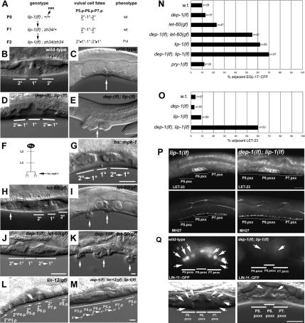
Protein phosphorylation by kinases and the subsequent dephosphorylation by phosphatases are key mechanisms that regulate intracellular signal transduction during development. Here, we report the identification of the receptor protein tyrosine phosphatase DEP-1 as a negative regulator of the Caenorhabditis elegans EGF receptor. DEP-1 amplifies in the developing vulva and the excretory system the small differences in the amount of EGF signal received by equivalent precursor cells to achieve binary cell fate decisions. During vulval development, DEP-1 inhibits EGFR signaling in the secondary cell lineage in parallel with the NOTCH-mediated lateral inhibition, while EGFR signaling simultaneously down-regulates DEP-1 and NOTCH expression in the primary cell lineage. This regulatory network of inhibitors results in the full activation of the EGFR/RAS/MAPK pathway in the primary vulval cells and at the same time keeps the EGFR/RAS/MAPK pathway inactive in the adjacent secondary cells. Mammalian Dep-1/Scc1 functions as a tumor-suppressor gene in the intestinal epithelium. Thus, mutations in human Dep-1 may promote tumor formation through a hyperactivation of the EGF receptor.


Figures

Figure 1.
Figure 1.
Identification and morphological characterization of dep-1. (A) Genetic screen to identify mutants with 2° cell fate transformations. Wild-type vulva in an L4 larva (B) and adult (C). (D) Vulva of a dep-1(lf); lip-1(lf) L4 larva. Note that the P5.p and P7.p descendants have detached from the cuticula and moved inward (9). (E) Adult dep-1(lf); lip-1(lf) animal exhibiting a protrusion of vulval tissue due to the mixed 1°/2° cell fate of P5.p and P7.p descendants. (F,G) Overexpression of MPK-1 after the first round of VPC cell divisions causes a phenotype similar to the dep-1(lf); lip-1(lf) L4 larva shown in D. (H) let-60 ras(gf) L4 larva. P5.p and P7.p have adopted the 2° fate, and their descendants remain attached to the cuticula, while some descendants of P4.p form an anterior pseudovulva (small arrow). (I) Adult let-60(gf) animal with a normal vulva in the center (large arrow) and an anterior pseudovulva (small arrow). A dep-1(lf); let-60(gf) L4 larva (J) and adult (K) animal exhibiting 2° cell fate transformations in addition to the Muv phenotype. (L) A lin-12 notch(gf) L4 larva, in which all VPCs have adopted the 2° fate. The orientations of the 2° fates are indicated with arrows. (M) A dep-1(lf); lin-12(gf); lip-1(lf) triple mutant displaying multiple 2° cell fate transformations. (N) Percentage of L3 larvae displaying egl-17::cfp expression in P5.px or P7.px cells. (O) Percentage of L3 larvae showing LET-23 expression in P5.p or P7.p descendants at the Pn.px and Pn.pxx stages. (P) LET-23 antibody staining of a lip-1(lf) and a dep-1(lf); lip-1(lf) mid-L3 larva at the Pn.pxx stage. MH27 staining of the adherens junctions in the animals is shown below. (Q) LIN-11::GFP expression from the syIs80 transgene (Gupta et al. 2003) in P5.p and P7.p descendants of wild-type and dep-1(lf); lip-1(lf) L4 larvae at the Pn.pxxx stage. Arrows point at the LIN-11::GFP expressing vulval cells. Bars, 10 μm.
Figure 2.
Figure 2.
Positional cloning of dep-1. (A) Map position, intron-exon structure, and fragments used for rescue and RNAi experiments. (B) Domain structure of DEP-1 (F44G4.8) compared with human DEP-1 and Drosophila Ptp4E. Open boxes represent the fibronectin-binding III repeats and black boxes the catalytic phosphatase domain. “I” indicates the percent sequence identity between the catalytic domains. The location of the zh34 stop mutation and the catalytic center are highlighted. (C) Dendogram showing the relationship between C. elegans (C.e.) DEP-1 (F44G4.8) and R-PTPs from Drosophila melanogaster (D.m.) and Homo sapiens (H.s.) calculated with the neighbor-joining method using the CLUSTAL X algorithm (Thompson et al. 1997). (D) Sequence alignment of the catalytic phosphatase domains of C. elegans DEP-1 with the catalytic domains of human class III R-PTPs. The asterisk indicates the position of the stop mutation in dep-1(zh34) animals, and the catalytic center containing the essential cysteine residue is underlined. GenBank accession numbers: (C.e.) DEP-1 CAA90125, (H.s.) DEP-1 Q12913, (H.s.) R-PTP-H NP002833, (H.s.) R-PTPβ P23467, (D.m.) PTP4E AAA76834, (D.m.) PTP10D P35992, (H.s.) R-PTP-RO AAH35960.
Figure 3.
Figure 3.
A substrate trapping mutant of DEP-1 binds LET-23 EGFR. (Left panel) Binding of LET-23 EGFR from N2 worm lysates to GST::DEP-1 fusion proteins detected on a Western blot with polyclonal LET-23 antibodies. Twenty percent of the amount of N2 lysate used in the binding reactions was loaded in the left-most lane. The band around 150 kDa corresponds to full-length LET-23. “n.s.” indicates a nonspecific cross-reacting band since a similar-sized band was detected in extracts from mammalian cells lacking LET-23 expression. The same amounts of recombinant proteins used for the binding reactions were loaded on a parallel gel stained with Coomassie blue shown in the right panel. For details on the assay conditions, see Materials and Methods.
Figure 4.
Figure 4.
DEP-1::GFP expression pattern during vulval development. (A-D) Expression pattern of the translational DEP-1::GFP reporter during vulval development. Note in C the lower levels of DEP::GFP in the two distal cells of the 2° lineages at the Pn.pxx stage. (E) Mutually exclusive DEP-1::GFP and LET-23 antibody staining at the Pn.pxx stage. (F) EGL-17::YFP expression at the Pn.pxx stage. Expression of the transcriptional dep-1p::gfp reporter in wild-type (G), lin-12(gf) (H), and lin-12(lf) (I) larvae at the Pn.pxx and Pn.px stages, respectively. Persisting DEP-1::GFP expression in all (J) or half (K) of the induced cells in sur-2(lf) mutants. Subcellular localization of DEP-1::GFP (L) and LET-23 (M) at the Pn.px stage. (N) Merged image. Arrows point at intracellular punctae containing both DEP-1::GFP and LET-23. Bars: N, 5 μm; otherwise, 10 μm. (O) Summary of the observed DEP-1::GFP expression pattern.
Figure 5.
Figure 5.
Cell-autonomous function of DEP-1 in the 2° cell lineage. Two examples of the informative mosaic animals. Arrows indicate the position of the nuclei of P5.p and P7.p descendants; arrowheads point at the nuclei of P6.p descendants. (A,C) Morphology of the P5.p and P7.p descendants at the L4 stage. (B,D) Expression of the SUR-5::GFP cell lineage marker indicating presence of the rescuing dep-1(+) array zhEx90. The insets show the upper focal planes where four of the eight P6.p descendants are located. (A,B) The animal from row 5 in Table 3. The descendants of P5.p lacked dep-1, while P6.p and P7.p descendants were wild type, resulting in a cell fate transformation of the P5.p descendants. (C,D) One of the three animals from row 7 in Table 3 in which the descendants of P6.p lacked dep-1, while P5.p and P7.p descendants were wild type. P5.p and P7.p adopted a normal 2° fate in this animal. Bar: D, 10 μm.
Figure 6.
Figure 6.
DEP-1 controls duct cell specification. (A) Lineage relationship between the duct cell precursors and phenotypes caused by let-60 ras mutations (Yochem et al. 1997). Nomarski (B) and GFP fluorescence (C) image of the duct cell in a wild-type L3 larva carrying the LIN-48::GFP reporter (Sewell et al. 2003). (D,E) let-60(gf) larva containing two duct cells. (F,G) dep-1(lf); lip-1(lf) animal exhibiting duct cell duplication. Arrows point at the nuclei of LIN-48::GFP-expressing cells. Bar, 5 μm. (H) Quantification of the duct cell duplications. “n” indicates the number of larvae scored for each genotype.
Figure 7.
Figure 7.
A model for DEP-1 function during vulval development. The inductive AC signal simultaneously down-regulates LIN-12 NOTCH and DEP-1 expression in P6.p via SUR-2, while LIN-12 and DEP-1 block the transduction of the inductive AC signal in P5.p and P7.p.

References

    1. Alonso A., Sasin, J., Bottini, N., Friedberg, I., Osterman, A., Godzik, A., Hunter, T., Dixon, J., and Mustelin, T. 2004. Protein tyrosine phosphatases in the human genome. Cell 117: 699-711. - PubMed
    1. Ambros V. 1999. Cell cycle-dependent sequencing of cell fate decisions in Caenorhabditis elegans vulva precursor cells. Development 126: 1947-1956. - PubMed
    1. Berset T., Hoier, E.F., Battu, G., Canevascini, S., and Hajnal, A. 2001. Notch inhibition of RAS signaling through MAP kinase phosphatase LIP-1 during C. elegans vulval development. Science 291: 1055-1058. - PubMed
    1. Bhaduri A. and Sowdhamini, R. 2003. A genome-wide survey of human tyrosine phosphatases. Protein Eng. 16: 881-888. - PubMed
    1. Blanchetot C., Chagnon, M., Dube, N., Halle, M., and Tremblay, M.L. 2005. Substrate-trapping techniques in the identification of cellular PTP targets. Methods 35: 44-53. - PubMed

Publication types