. 2005 Jun;187(11):3721-30.
doi: 10.1128/JB.187.11.3721-3730.2005.

Expression and site-directed mutagenesis of the lactococcal abortive phage infection protein AbiK

Affiliations

Expression and site-directed mutagenesis of the lactococcal abortive phage infection protein AbiK

Louis-Charles Fortier et al. J Bacteriol. 2005 Jun.

Abstract

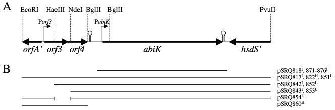
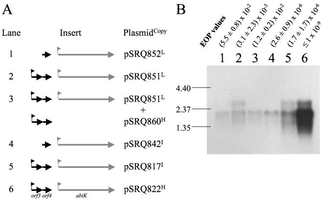
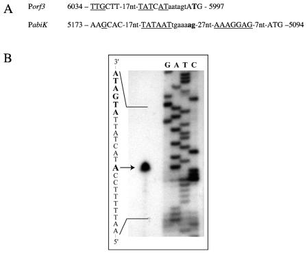
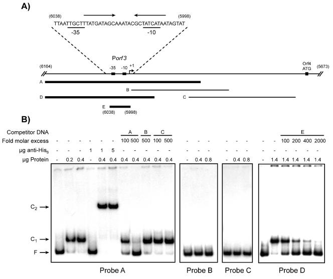
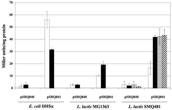
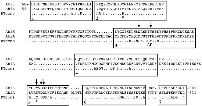
Abortive infection mechanisms of Lactococcus lactis form a heterogeneous group of phage resistance systems that act after early phage gene expression. One of these systems, AbiK, aborts infection of the three most prevalent lactococcal phage groups of the dairy industry. In this study, it is demonstrated that the antiphage activity depends on the level of expression of the abiK gene and on the presence of a reverse transcriptase (RT) motif in AbiK. The abiK gene was shown to be part of an operon that includes two additional open reading frames, with one of these encoding a phage-related transcriptional repressor named Orf4. Expression of AbiK is driven by two promoters, PabiK and Porf3, the latter being repressed by Orf4 in vivo. Binding of the purified Orf4 to the Porf3 promoter was demonstrated in vitro by gel retardation assays. The N-terminal half of the deduced AbiK protein possesses an RT motif that was modified by site-directed mutagenesis. Conservative mutations in key positions resulted in the complete loss of the resistance phenotype. These data suggest that an RT activity might be involved in the phage resistance activity of AbiK. A model for the mode of action of AbiK is proposed.


Figures

FIG. 1.
FIG. 1.
Genetic organization of the DNA fragment of pSRQ800 that encodes phage resistance. Panel A: open reading frames, promoters (bent arrows), and rho-independent terminators (stem-loops) identified by bioinformatics analyses. Panel B: representation of the DNA fragments cloned into the different plasmids used in this study. The superscript letter at the end of the plasmid name represents the plasmid copy number in L. lactis (H = high; I = intermediate; L = low).
FIG. 2.
FIG. 2.
Transcription of the abiK operon. Panel A shows the plasmids carried by the six strains used, along with a schematic representation of the cloned insert. The corresponding plasmid copy number in L. lactis MG1363 is indicated by a superscript letter at the end of the plasmid name (see the legend to Fig. 1). The pNZ123 vector was cointroduced as a control in addition to the indicated plasmids in lanes 1 and 2. Panel B presents the Northern blot analysis with a probe corresponding to the abiK gene. To facilitate the comparison of the different constructs, some EOP values from Table 2 (without pSRQ860) are shown above the blot. Bands of the molecular weight marker (in kbp) are indicated on the left.
FIG. 3.
FIG. 3.
Promoter determination. Panel A: DNA sequence and transcription initiation sites (in boldface) of the Porf3 and PabiK promoters. The −35 and −10 regions, the ribosome binding site of abiK, and the start codons of orf3 and abiK are in capital letters. Nucleic acids that are identical to the consensus sequence are underlined. The coordinates correspond to the DNA sequence of pSRQ800 (accession no. U35629). Panel B: primer extension analysis of the Porf3 promoter. Total RNA from an L. lactis MG1363 strain carrying the pSRQ817 plasmid was purified, and primer extension was performed as described in Materials and Methods. The lower-strand DNA sequence shown on the left was obtained with the same primer in a T7-based sequencing reaction. The −10 box is highlighted in boldface. The transcription initiation start site is indicated by an arrow.
FIG. 4.
FIG. 4.
Gel retardation assays (EMSA) with recombinant Orf4. Panel A shows the different fragments (indicated by letters A to E) used as labeled probes (10,000 cpm/reaction) or unlabeled competitor DNA. Thick lines represent the fragments retarded by Orf4 in EMSA, whereas thin lines represent unbound fragments. The DNA sequence corresponding to fragment E is shown above, along with the −10 and −35 boxes (underlined characters). The inverted repeats are indicated by arrows above the sequence. Some of the coordinates are shown in parentheses and correspond to the pSRQ800 sequence (GenBank accession no. U35629). The transcription initiation start site (+1) is indicated by a bent arrow. Panel B, EMSA with the various fragments shown in panel A. Unlabeled competitor DNA fragments are indicated by letters (corresponding to fragments in panel A). F. free probe; C1, Orf4-DNA complex; C2, anti-His6-Orf4-DNA complex.
FIG. 5.
FIG. 5.
β-Galactosidase activity in various E. coli and L. lactis strains at 37°C (white bars), at 30°C (black bars), and during infection by phage ul36 (horizontally striped bars, after 15 min; diagonally striped bars, after 30 min). These data represent the means and standard deviations from at least three experiments.
FIG. 6.
FIG. 6.
Alignment of AbiK, AbiA, and the RT motifs of Xiong and Eickbush (70). The identical residues in the amino acid sequences of AbiK and AbiA are in boldface. h, hydrophobic residue; p, small polar residue; c, charged residue. The amino acids that were replaced by site-directed mutagenesis are identified by arrows.

References

    1. Avidan, O., M. E. Meer, I. Oz, and A. Hizi. 2002. The processivity and fidelity of DNA synthesis exhibited by the reverse transcriptase of bovine leukemia virus. Eur. J. Biochem. 269:859-867. - PubMed
    1. Baichoo, N., and J. D. Helmann. 2002. Recognition of DNA by Fur: a reinterpretation of the Fur box consensus sequence. J. Bacteriol. 184:5826-5832. - PMC - PubMed
    1. Bakhanashvili, M., O. Avidan, and A. Hizi. 1996. Mutational studies of human immunodeficiency virus type 1 reverse transcriptase: the involvement of residues 183 and 184 in the fidelity of DNA synthesis. FEBS Lett. 391:257-262. - PubMed
    1. Bidnenko, E., S. D. Ehrlich, and M. C. Chopin. 1998. Lactococcus lactis phage operon coding for an endonuclease homologous to RuvC. Mol. Microbiol. 28:823-834. - PubMed
    1. Birnboim, H. C. 1983. A rapid alkaline extraction method for the isolation of plasmid DNA. Methods Enzymol. 100:243-255. - PubMed

Publication types

MeSH terms

Substances