. 2005 Jun;187(11):3779-85.
doi: 10.1128/JB.187.11.3779-3785.2005.

Role of pi dimers in coupling ("handcuffing") of plasmid R6K's gamma ori iterons

Affiliations

Role of pi dimers in coupling ("handcuffing") of plasmid R6K's gamma ori iterons

Selvi Kunnimalaiyaan et al. J Bacteriol. 2005 Jun.

Abstract

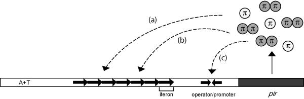
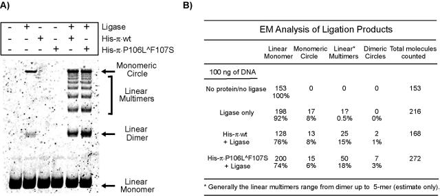
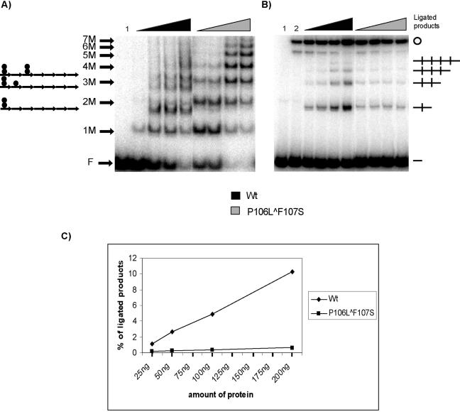
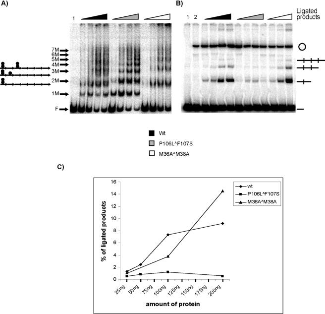
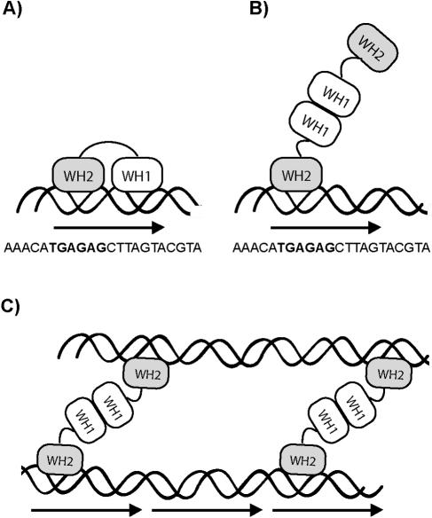
One proposed mechanism of replication inhibition in iteron-containing plasmids (ICPs) is "handcuffing," in which the coupling of origins via iteron-bound replication initiator (Rep) protein turns off origin function. In minimal R6K replicons, copy number control requires the interaction of plasmid-encoded pi protein with the seven 22-bp iterons of the gamma origin of replication. Like other related Rep proteins, pi exists as both monomers and dimers. However, the ability of pi dimers to bind iterons distinguishes R6K from most other ICPs, where only monomers have been observed to bind iterons. Here, we describe experiments to determine if monomers or dimers of pi protein are involved in the formation of handcuffed complexes. Standard ligation enhancement assays were done using pi variants with different propensities to bind iterons as monomers or dimers. Consistent with observations from several ICPs, a hyperreplicative variant (pi.P106L(wedge)F107S) exhibits deficiencies in handcuffing. Additionally, a novel dimer-biased variant of pi protein (pi.M36A(wedge)M38A), which lacks initiator function, handcuffs iteron-containing DNA more efficiently than does wild-type pi. The data suggest that pi dimers mediate handcuffing, supporting our previously proposed model of handcuffing in the gamma ori system. Thus, dimers of pi appear to possess three distinct inhibitory functions with respect to R6K replication: transcriptional autorepression of pi expression, in cis competition (for origin binding) with monomeric activator pi, and handcuffing-mediated inhibition of replication in trans.


Figures

FIG. 1.
FIG. 1.
Roles of π binding to γ ori. The pir gene encodes π protein, which exists in two forms: monomers (white) and dimers (gray). The seven direct repeats of γ ori, also called iterons, are indicated by tandem arrows, whereas inverted arrows represent the inverted repeat in the pir gene operator/promoter. π is a multifunctional protein: monomers activate replication (a), dimers inhibit replication (b), and dimers autorepress pir transcription (c).
FIG. 2.
FIG. 2.
Detection of π-dependent handcuffing in the presence of excess DNA. (A) One hundred nanograms of EcoRV fragment containing seven DRs was incubated in the presence or absence of 125 ng of His-π·wt or His-π·P106LF107S and with or without 0.5 unit of ligase as indicated. The samples were processed as described in Materials and Methods. Linear DNA monomers, linear dimers, and monomeric circles are indicated by arrows; linear DNA multimers are indicated by a bracket. (B) Categorization of ligation products by electron microscopy.
FIG. 3.
FIG. 3.
Binding of π to the iteron and detection of π-dependent handcuffing by ligation enhancement under limiting DNA conditions. A 0.5-ng amount of 32P-end-labeled EcoRV fragment containing seven DRs was utilized, along with increasing amounts of His-π·wt and His-π· P106LF107S. (A) Hypothetical representation of π monomers and π dimers bound to the seven iterons in EMSA is shown (left). F indicates free DNA; 1 M to 7 M indicates the number of π monomer-bound iterons (e.g., 7 M indicates that the seven-DR fragment has each iteron filled by a monomer). Triangles indicate increasing amounts of protein added to the reaction mixtures: 25, 50, 100, and 200 ng. Lane 1 contains DNA without protein. (B) The remaining samples were treated with 0.5 unit of ligase and processed as described in Materials and Methods. Lane 1 contains DNA only (without protein or ligase); lane 2 contains DNA with 0.5 unit of ligase. Handcuffed products (linear DNA dimer, linear trimer, linear tetramer, and monomeric circles) and free DNA (—) are indicated. (C) Quantification of ligated products; results are the averages from three independent experiments.
FIG. 4.
FIG. 4.
Detection of handcuffing using wt and copy-up and dimeric variants of π. A 0.5-ng amount of 32P-labeled EcoRV fragment containing seven DRs was utilized in this experiment with increasing amounts of His-π·wt, His-π·P106LF107S, or His-π·M36AM38A. (A) Hypothetical representation of π monomers and π dimers bound to the seven iterons in EMSA is shown (left). F indicates free DNA; 1 M to 7 M indicates the number of π monomer-bound iterons (e.g., 7 M indicates that the seven-DR fragment has each iteron filled by a monomer). Triangles indicate increasing amounts of protein added to the reaction mixtures: 25, 50, 100, and 200 ng. (B) The remaining samples were treated with 0.5 unit of ligase and processed as described in Materials and Methods. For the sample containing His-π·M36AM38A, only 25-, 100-, and 200-ng samples were analyzed. Handcuffed products (linear DNA dimer, linear trimer, linear tetramer, and monomeric circles) and free DNA (—) are indicated. (C) Quantification of ligated products.
FIG. 5.
FIG. 5.
A schematic representation of π bound to iterons. π protein is indicated by two oval winged helix domains connected by a linker, paired wavy lines indicate double-stranded DNA, and an arrow indicates a single iteron, the sequence of which is shown. (A) π monomer bound to a single iteron using both WH1 and WH2 domains. (B) π dimer bound to a single iteron using only the WH2 domain. (C) Proposed model for handcuffing in which both WH2 domains of preformed π dimers contact two iteron-containing DNA fragments. The figure is not drawn to scale.

Similar articles

Cited by

References

    1. Abhyankar, M. M., J. M. Reddy, R. Sharma, E. Bullesbach, and D. Bastia. 2004. Biochemical investigations of control of replication initiation of plasmid R6K. J. Biol. Chem. 279:6711-6719. - PubMed
    1. Blasina, A., B. L. Kittell, A. E. Toukdarian, and D. R. Helinski. 1996. Copy-up mutants of the plasmid RK2 replication initiation protein are defective in coupling RK2 replication origins. Proc. Natl. Acad. Sci. USA 93:3559-3564. - PMC - PubMed
    1. Chattoraj, D. K., and T. D. Schneider. 1997. Replication control of plasmid P1 and its host chromosome: the common ground. Prog. Nucleic Acid Res. Mol. Biol. 57:145-186. - PubMed
    1. Das, N., and D. K. Chattoraj. 2004. Origin pairing (‘handcuffing’) and unpairing in the control of P1 plasmid replication. Mol. Microbiol. 54:836-849. - PubMed
    1. del Solar, G., R. Giraldo, M. J. Ruiz-Echevarría, M. Espinosa, and R. Díaz-Orejas. 1998. Replication and control of circular bacterial plasmids. Microbiol. Mol. Biol. Rev. 62:434-464. - PMC - PubMed

Publication types

MeSH terms