The ExsA protein of Bacillus cereus is required for assembly of coat and exosporium onto the spore surface
- PMID: 15901704
- PMCID: PMC1112046
- DOI: 10.1128/JB.187.11.3800-3806.2005
The ExsA protein of Bacillus cereus is required for assembly of coat and exosporium onto the spore surface
Abstract
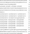
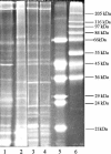
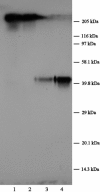
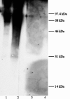
The outermost layer of spores of the Bacillus cereus family is a loose structure known as the exosporium. Spores of a library of Tn917-LTV1 transposon insertion mutants of B. cereus ATCC 10876 were partitioned into hexadecane; a less hydrophobic mutant that was isolated contained an insertion in the exsA promoter region. ExsA is the equivalent of SafA (YrbA) of Bacillus subtilis, which is also implicated in spore coat assembly; the gene organizations around both are identical, and both proteins contain a very conserved N-terminal cortex-binding domain of ca. 50 residues, although the rest of the sequence is much less conserved. In particular, unlike SafA, the ExsA protein contains multiple tandem oligopeptide repeats and is therefore likely to have an extended structure. The exsA gene is expressed in the mother cell during sporulation. Spores of an exsA mutant are extremely permeable to lysozyme and are blocked in late stages of germination, which require coat-associated functions. Two mutants expressing differently truncated versions of ExsA were constructed, and they showed the same gross defects in the attachment of exosporium and spore coat layers. The protein profile of the residual exosporium harvested from spores of the three mutants--two expressing truncated proteins and the mutant with the original transposon insertion in the promoter region--showed some differences from the wild type and from each other, but the major exosporium glycoproteins were retained. The exsA gene is extremely important for the normal assembly and anchoring of both the spore coat and exosporium layers in spores of B. cereus.
Figures


Similar articles
-
ExsY and CotY are required for the correct assembly of the exosporium and spore coat of Bacillus cereus.J Bacteriol. 2006 Nov;188(22):7905-13. doi: 10.1128/JB.00997-06. Epub 2006 Sep 15. J Bacteriol. 2006. PMID: 16980471 Free PMC article.
-
Sporulation Temperature Reveals a Requirement for CotE in the Assembly of both the Coat and Exosporium Layers of Bacillus cereus Spores.Appl Environ Microbiol. 2015 Oct 23;82(1):232-43. doi: 10.1128/AEM.02626-15. Print 2016 Jan 1. Appl Environ Microbiol. 2015. PMID: 26497467 Free PMC article.
-
Genes of Bacillus cereus and Bacillus anthracis encoding proteins of the exosporium.J Bacteriol. 2003 Jun;185(11):3373-8. doi: 10.1128/JB.185.11.3373-3378.2003. J Bacteriol. 2003. PMID: 12754235 Free PMC article.
-
Structure, assembly, and function of the spore surface layers.Annu Rev Microbiol. 2007;61:555-88. doi: 10.1146/annurev.micro.61.080706.093224. Annu Rev Microbiol. 2007. PMID: 18035610 Review.
-
The Exosporium Layer of Bacterial Spores: a Connection to the Environment and the Infected Host.Microbiol Mol Biol Rev. 2015 Dec;79(4):437-57. doi: 10.1128/MMBR.00050-15. Microbiol Mol Biol Rev. 2015. PMID: 26512126 Free PMC article. Review.
Cited by
-
A novel spore protein, ExsM, regulates formation of the exosporium in Bacillus cereus and Bacillus anthracis and affects spore size and shape.J Bacteriol. 2010 Aug;192(15):4012-21. doi: 10.1128/JB.00197-10. Epub 2010 Jun 11. J Bacteriol. 2010. PMID: 20543075 Free PMC article.
-
Localization and assembly of the novel exosporium protein BetA of Bacillus anthracis.J Bacteriol. 2011 Oct;193(19):5098-104. doi: 10.1128/JB.05658-11. Epub 2011 Aug 5. J Bacteriol. 2011. PMID: 21821770 Free PMC article.
-
Clostridium difficile spore biology: sporulation, germination, and spore structural proteins.Trends Microbiol. 2014 Jul;22(7):406-16. doi: 10.1016/j.tim.2014.04.003. Epub 2014 May 7. Trends Microbiol. 2014. PMID: 24814671 Free PMC article. Review.
-
Bacillus anthracis exosporium protein BclA affects spore germination, interaction with extracellular matrix proteins, and hydrophobicity.Infect Immun. 2007 Nov;75(11):5233-9. doi: 10.1128/IAI.00660-07. Epub 2007 Aug 20. Infect Immun. 2007. PMID: 17709408 Free PMC article.
-
The Bacillus cereus GerN and GerT protein homologs have distinct roles in spore germination and outgrowth, respectively.J Bacteriol. 2008 Sep;190(18):6148-52. doi: 10.1128/JB.00789-08. Epub 2008 Jul 18. J Bacteriol. 2008. PMID: 18641133 Free PMC article.
References
-
- Barlass, P. J., C. W. Houston, M. O. Clements, and A. Moir. 2002. Germination of Bacillus cereus spores in response to l-alanine and to inosine: the roles of gerL and gerQ operons. Microbiology 148:2089-2095. - PubMed
-
- Boland, F. M., A. Atrih, H. Chirakkal, S. J. Foster, and A. Moir. 2000. Complete spore-cortex hydrolysis during germination of Bacillus subtilis 168 requires SleB and YpeB. Microbiology 146:57-64. - PubMed
-
- Bone, E. J., and D. J. Ellar. 1989. Transformation of Bacillus thuringiensis by electroporation. FEMS Lett. 58:171-178. - PubMed
Publication types
MeSH terms
Substances
Associated data
- Actions
LinkOut - more resources
Full Text Sources
Other Literature Sources
Molecular Biology Databases

