. 2005 Jun;187(11):3848-54.
doi: 10.1128/JB.187.11.3848-3854.2005.

Structures of the iron-sulfur flavoproteins from Methanosarcina thermophila and Archaeoglobus fulgidus

Affiliations

Structures of the iron-sulfur flavoproteins from Methanosarcina thermophila and Archaeoglobus fulgidus

Susana L A Andrade et al. J Bacteriol. 2005 Jun.

Abstract

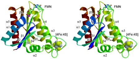
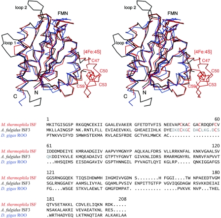
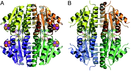
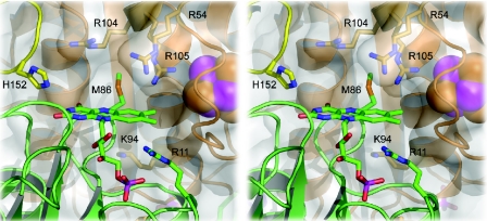
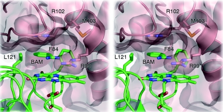
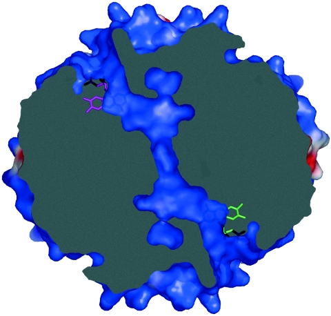
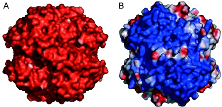
Iron-sulfur flavoproteins (ISF) constitute a widespread family of redox-active proteins in anaerobic prokaryotes. Based on sequence homologies, their overall structure is expected to be similar to that of flavodoxins, but in addition to a flavin mononucleotide cofactor they also contain a cubane-type [4Fe:4S] cluster. In order to gain further insight into the function and properties of ISF, the three-dimensional structures of two ISF homologs, one from the thermophilic methanogen Methanosarcina thermophila and one from the hyperthermophilic sulfate-reducing archaeon Archaeoglobus fulgidus, were determined. The structures indicate that ISF assembles to form a tetramer and that electron transfer between the two types of redox cofactors requires oligomerization to juxtapose the flavin mononucleotide and [4Fe:4S] cluster bound to different subunits. This is only possible between different monomers upon oligomerization. Fundamental differences in the surface properties of the two ISF homologs underscore the diversity encountered within this protein family.


Figures

FIG. 1.
FIG. 1.
Stereo view of the iron-sulfur flavoprotein monomer from M. thermophila, with colors ranging from blue at the N terminus to red at the C terminus. Secondary structure elements are labeled according to their order of appearance along the peptide chain. Within a single ISF monomer, the shortest distance between the FMN cofactor and the iron-sulfur cluster is ∼24 Å.
FIG. 2.
FIG. 2.
Structural alignment of ISF-Mt (red) and the iron-free variant of ISF3-Af (black) with the C-terminal, flavodoxin-like domain of D. gigas ROO (blue). In the ISF-Mt structure, the four cysteine residues that coordinate the iron-sulfur cluster are labeled. Two protruding loops, labeled loop 1 and loop 2, are observed in the A. fulgidus structure. Their positions suggest involvement in stabilization of the tetrameric form of ISF. In the ROO domain, the cluster-binding loop is absent, but the overall fold is highly similar to that in ISF.
FIG. 3.
FIG. 3.
Tetramers of M. thermophila ISF (A) and A. fulgidus ISF3 (B). In panel B, the loop connecting the β2 sheet with helix α2 is disordered in the absence of the metal cluster.
FIG. 4.
FIG. 4.
Detail of the flavin site of M. thermophila ISF. Although the FMN moiety is surrounded by a number of positively charged residues, the effective electrostatic surface potential in this region is negative (see Fig. 7).
FIG. 5.
FIG. 5.
Flavin site of A. fulgidus ISF3. In contrast to ISF-Mt, access to this site is limited due to the side chain of Arg99 from a neighboring monomer. Stacked between the flavin and a cation-π interaction with the side chain of Arg102, a benzamidine molecule (BAM) from the crystallization buffer was modeled hydrogen bonded to Arg99. The aromatic ring of the benzamidine molecule is situated in a hydrophobic pocket created by Leu121 and Phe84. The benzamidine sits at the entrance of a channel that crosses the entire ISF3 tetramer (see Fig. 6).
FIG. 6.
FIG. 6.
A channel crosses the tetramer of A. fulgidus ISF3, connecting the FMN molecules of two opposing monomers. A second channel is found connecting the other two, symmetry-equivalent FMN molecules (not shown). The image shows a slice through the ISF3-Af tetramer, as shown in Fig. 7B, with identical surface coloring according to the electrostatic surface potential.
FIG. 7.
FIG. 7.
Electrostatic surface potentials for the tetramers of M. thermophila ISF (A) and A. fulgidus ISF3 (B), oriented as shown in Fig. 3. Potentials were calculated with the program DELPHI, using dielectric values (ɛ) of 4 for the protein interior and 80 for the surrounding solvent. To allow a more realistic comparison of surface properties, the cluster-binding loop of ISF-Mt was grafted onto the model of ISF3-Af, and the diverging amino acid side chains were replaced. Potentials are contoured from −75 kT (red) to 75 kT (blue).

References

    1. Becker, D. F., U. Leartsakulpanich, K. K. Surerus, J. G. Ferry, and S. W. Ragsdale. 1998. Electrochemical and spectroscopic properties of the iron-sulfur flavoprotein from Methanosarcina thermophila. J. Biol. Chem. 273:26462-26469. - PubMed
    1. Brünger, A. T., P. D. Adams, G. M. Clore, W. L. Delano, P. Gros, R. W. Grosse Kunstleve, J. S. Jiang, J. Kuszewski, M. Nilges, N. S. Pannu, R. J. Read, L. M. Rice, T. Simonson, and G. L. Warren. 1998. Crystallography and NMR system: a new software suite for macromolecular structure determination. Acta Crystallogr. Sect. D 54:905-921. - PubMed
    1. Collaborative Computational Project No. 4. 1994. The CCP4 Suite: programs for protein crystallography. Acta Crystallogr. Ser. D 50:760-763. - PubMed
    1. Darnault, C., A. Volbeda, E. J. Kim, P. Legrand, X. Vernede, P. A. Lindahl, and J. C. Fontecilla-Camps. 2003. Ni-Zn-[Fe4-S4] and Ni-Ni-[Fe4-S4] clusters in closed and open subunits of acetyl-CoA synthase/carbon monoxide dehydrogenase. Nat. Struct. Biol. 10:271-279. - PubMed
    1. Ding, Y. H. R., and J. G. Ferry. 2004. Flavin mononucleotide-binding flavoprotein family in the domain Archaea. J. Bacteriol. 186:90-97. - PMC - PubMed

Publication types

MeSH terms