Comparative Study
. 2005 Sep;94(3):1814-24.
doi: 10.1152/jn.00374.2005. Epub 2005 May 18.

Synaptic transmission at the cochlear nucleus endbulb synapse during age-related hearing loss in mice

Affiliations
Comparative Study

Synaptic transmission at the cochlear nucleus endbulb synapse during age-related hearing loss in mice

Yong Wang et al. J Neurophysiol. 2005 Sep.

Abstract

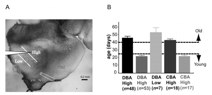
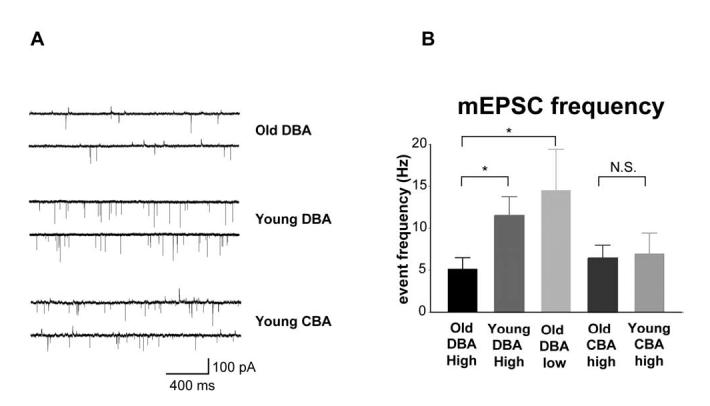
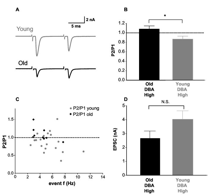
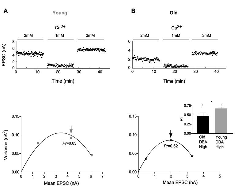
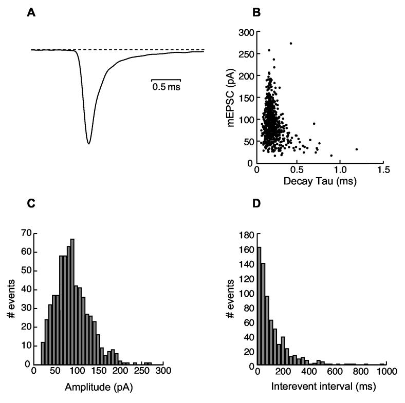
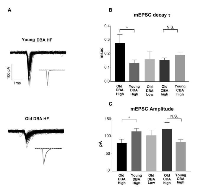
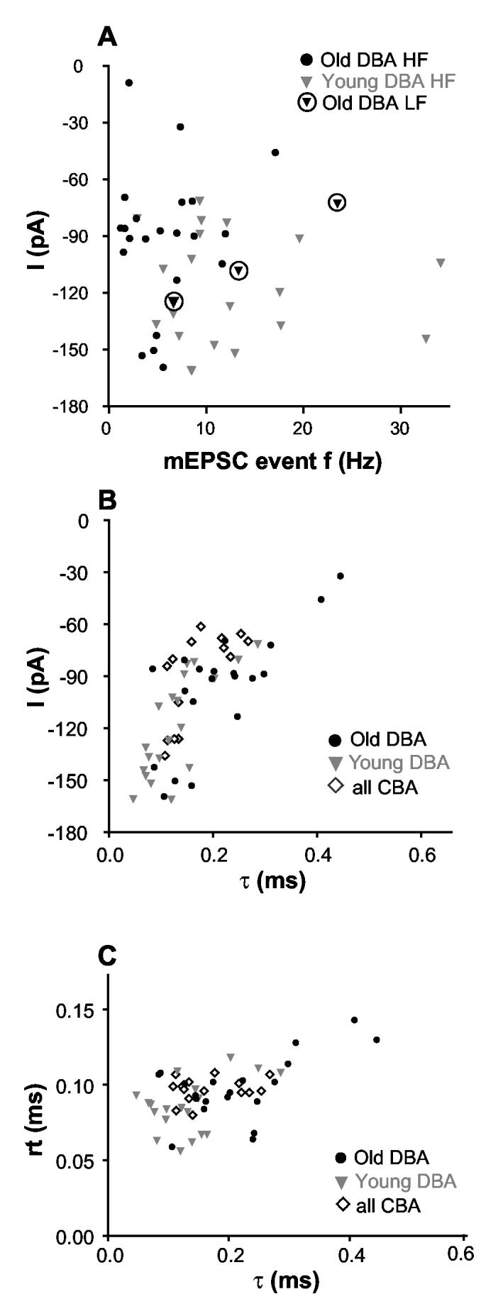
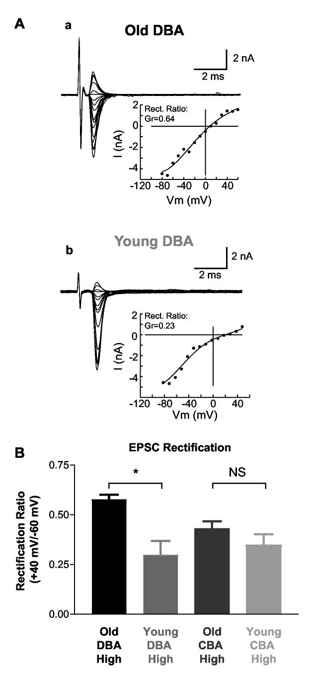
Age-related hearing loss (AHL) typically starts from high-frequency regions of the cochlea and over time invades lower-frequency regions. During this progressive hearing loss, sound-evoked activity in spiral ganglion cells is reduced. DBA mice have an early onset of AHL. In this study, we examined synaptic transmission at the endbulb of Held synapse between auditory nerve fibers and bushy cells in the anterior ventral cochlear nucleus (AVCN). Synaptic transmission in hearing-impaired high-frequency areas of the AVCN was altered in old DBA mice. The spontaneous miniature excitatory postsynaptic current (mEPSC) frequency was substantially reduced (about 60%), and mEPSCs were significantly slower (about 115%) and smaller (about 70%) in high-frequency regions of old (average age 45 days) DBA mice compared with tonotopically matched regions of young (average age 22 days) DBA mice. Moreover, synaptic release probability was about 30% higher in high-frequency regions of young DBA than that in old DBA mice. Auditory nerve-evoked EPSCs showed less rectification in old DBA mice, suggesting recruitment of GluR2 subunits into the AMPA receptor complex. No similar age-related changes in synaptic release or EPSCs were found in age-matched, normal hearing young and old CBA mice. Taken together, our results suggest that auditory nerve activity plays a critical role in maintaining normal synaptic function at the endbulb of Held synapse after the onset of hearing. Auditory nerve activity regulates both presynaptic (release probability) and postsynaptic (receptor composition and kinetics) function at the endbulb synapse after the onset of hearing.


Figures

Fig. 1
Fig. 1
Illustration of frequency regions where electrophysiology recordings were made. A) A 200-250 μm thick parasagittal slice of brainstem containing cochlear nucleus was cut. The AVCN was divided along the isofrequency line into three equal zones representing high, medium and low frequency regions. There was no overlap between the high and low frequency areas. The placement of the bipolar stimulating for electrode auditory nerve fiber stimulation was indicated. B) Experimental animal ages and tonotopic regions where cells were recorded. Animals younger than 25 days were considered to be young, whereas animals older than 40 days were considered old.
Fig. 2
Fig. 2
Spontaneous mEPSC event frequency was reduced in hearing-impaired old DBA mice. A) Representative mEPSC traces from high frequency bushy cells. Cells were held near their resting membrane potential at -60mV. No glycine receptor antagonist was included in the bathing solution. B) Spontaneous mEPSC event frequency from different age and frequency groups. mEPSC event frequencies from high frequency regions were lower in old DBA mice than that in young DBA mice, whereas the event frequency for normal hearing low frequency old DBA mice was similar to that of young DBA mice. mEPSC event frequency was not significantly different between old and young CBA high frequency bushy cells.
Fig. 3
Fig. 3
Release probability at endbulb synapses was lower in hearing-impaired DBA mice. A) Sample paired-pulse responses from HF bushy cells in young and old DBA mice. A paired-pulse protocol was used to measure the initial release probability at the endbulb synapse. B) The P2/P1 ratios were significantly different between young and old DBA mice. C) Paired-pulse ratios from all individual cells were plotted against the spontaneous mEPSC event frequency. Cells with strong paired-pulse depression tended to have higher mEPSC event frequency. D) The evoked EPSC amplitude in old and young DBA mice showed no statistical difference although there was a tendency for old animals to have smaller EPSC amplitude.
Fig. 4
Fig. 4
Mean-variance analysis of release probability in high-frequency cells of DBA mice. A) Evoked EPSCs recorded from a high frequency bushy cell in a young DBA mouse. Stable EPSCs were measured at varying extracellular calcium concentrations (top panel) and plotted against variance (bottom panel). A parabola was fitted (see Methods) to estimate release probability (Pr). The arrow indicates the Pr estimate in physiological calcium (2 mM). B) EPSCs recorded from an old DBA mouse. Inset shows summary data for Pr at 2 mM [Ca] from high frequency bushy cells in young and old DBA mice.
Fig. 5
Fig. 5
Properties of spontaneous miniature EPSCs. A) The normalized average of all the mEPSCs (∼500 events) recorded from a high frequency bushy cell in a young DBA mouse for 40 seconds. B) The amplitudes of all individual events were plotted against their respective decay time constants. Although there was a broad range of mEPSC amplitude size, for a given cell, the majority of mini EPSCs had similar decay time constant. C) The distribution pattern of all mEPSC amplitudes. D) mEPSC event intervals showed a Poisson distribution with a CV=1.05.
Fig. 6
Fig. 6
Spontaneous mEPSCs had slower decay time constant in hearing-impaired DBA bushy cells. A) Sample mEPSCs recorded from 2 bushy cells in HF regions from a young and an old DBA mouse. All detected mEPSCs were aligned to their onset. Insets: normalized average of the mEPSCs superimposed with the first order exponential decay (dark trace). B) Decay time constants for young and old bushy cells in high frequency regions of DBA mice were significantly different. Decay time constants for all cells from normal hearing regions of the AVCN were comparable between old DBA and young CBA HF as well as old CBA HF cells. C) Spontaneous mEPSC amplitude was significantly different between hearing-impaired old DBA mice and young DBA mice. There was no statistical difference between the normal hearing low frequency old DBA and the high frequency young DBA cells, nor between the old CBA HF and the young CBA HF cells.
Fig. 7
Fig. 7
mEPSCs from cells not affected by hearing loss tended to be larger, more frequent, and had faster decay constant. A) mEPSC amplitude was plotted against event frequency for all individual bushy cells recorded from DBA mice. B) Larger mEPSCs tended to have faster decay time course (r2=0.72). mEPSC amplitude and decay time constant were plotted from bushy cells in the high frequency region of DBA and CBA of all ages. There were no apparent differences for amplitude/τ relationship between the groups. C) mEPSCs with a slow decay τ did not have a slow rising time. The rising and decay time constants of mEPSCs of all individual cells are plotted.
Fig. 8
Fig. 8
AMPA receptor mediated currents had less rectification in high frequency cells of impaired hearing old DBA mice. A) EPSC current-voltage relationships from representative individual bushy cells from old and young DBA mice are plotted. B) Comparison of the rectification index in both DBA and CBA mice. The rectification index was computed by taking the ratio of conductance of the cell at +40 mV and -60 mV; smaller numbers indicate greater rectification. Evoked EPSCs at various holding voltages were recorded with electrodes containing 50μM NHPP-spermine. In high frequency cells of old DBA mice, EPSC rectification was significantly weaker than that in young animals. No difference was observed between age-matched groups in CBA mice, nor between young DBA and CBA mice of either age group.

References

    1. Bellingham MC, Lim R, Walmsley B. Developmental changes in EPSC quantal size and quantal content at a central glutamatergic synapse in rat. J Physiol. 1998;511:861–869. - PMC - PubMed
    1. Berglund AM, Brown MC. Central trajectories of type II spiral ganglion cells from various cochlear regions in mice. Hear Res. 1994;75:121–130. - PubMed
    1. Boulter J, Hollmann M, O’Shea-Greenfield A, Hartley M, Deneris E, Maron C, Heinemann S. Molecular cloning and functional expression of glutamate receptor subunit genes. Science. 1990;249:1033–1037. - PubMed
    1. Bowie D, Mayer ML. Inward rectification of both AMPA and kainate subtype glutamate receptors generated by polyamine-mediated ion channel block. Neuron. 1995;15:453–462. - PubMed
    1. Brawer JR, Morest DK, Kane EC. The neuronal architecture of the cochlear nucleus of the cat. J Comp Neurol. 1974;155:251–300. - PubMed

Publication types

MeSH terms