Comparative Study
. 2005 May 18;25(20):4889-97.
doi: 10.1523/JNEUROSCI.0679-05.2005.

Mutually exclusive glomerular innervation by two distinct types of olfactory sensory neurons revealed in transgenic zebrafish

Affiliations
Comparative Study

Mutually exclusive glomerular innervation by two distinct types of olfactory sensory neurons revealed in transgenic zebrafish

Yuki Sato et al. J Neurosci. .

Abstract

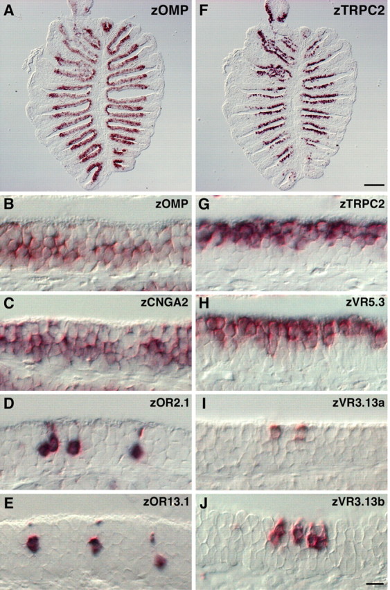
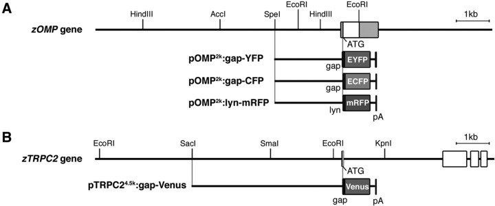
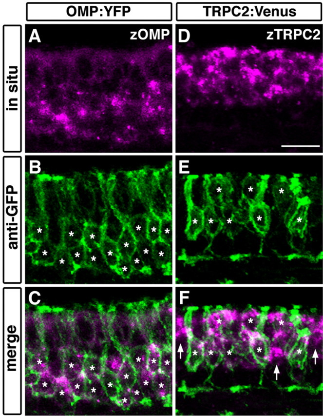
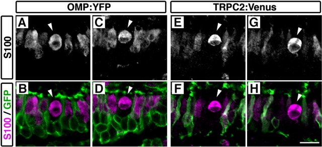
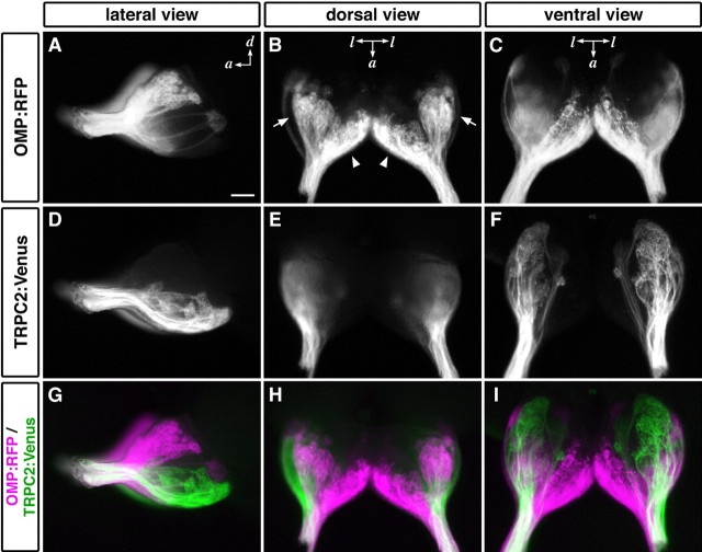
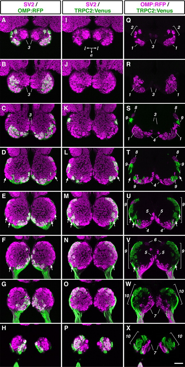
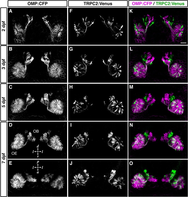
The olfactory epithelium of fish contains two major types of olfactory sensory neurons (OSNs) that are distinct morphologically (ciliated vs microvillous) and possibly functionally. Here, we found that these OSNs express different sets of signal transduction machineries: the ciliated OSNs express OR-type odorant receptors, cyclic nucleotide-gated channel A2 subunit, and olfactory marker protein (OMP), whereas the microvillous OSNs express V2R-type receptors and transient receptor potential channel C2 (TRPC2). To visualize patterns of axonal projection from the two types of OSNs to the olfactory bulb (OB), we generated transgenic zebrafish in which spectrally distinct fluorescent proteins are expressed in the ciliated and microvillous OSNs under the control of OMP and TRPC2 gene promoters, respectively. An observation of whole-mount OB in adult double-transgenic zebrafish revealed that the ciliated OSNs project axons mostly to the dorsal and medial regions of the OB, whereas the microvillous OSNs project axons to the lateral region of the OB. A careful histological examination of OB sections clarified that the axons from the two distinct types of OSNs target different glomeruli in a mutually exclusive manner. This segregation is already established at very early developmental stages in zebrafish embryos. These findings clearly demonstrate the relationships among cell morphology, molecular signatures, and axonal terminations of the two distinct types of OSNs and suggest that the two segregated neural pathways are responsible for coding and processing of different types of odor information in the zebrafish olfactory system.


Figures

Figure 1.
Figure 1.
Two distinct types of OSNs with different molecular signatures in zebrafish OE. A-J, RNA in situ hybridization was performed on horizontal sections of the zebrafish olfactory rosette using digoxigenin-labeled probes specific for the following molecules: zOMP (A, B), zCNGA2 (C), zOR2.1 (D), zOR13.1 (E), zTRPC2 (F, G), zVR5.3 (H), zVR3.13a (I), and zVR3.13b (J). Note that zOMP (B), zCNGA2 (C), and OR-type receptors (D, E) were expressed in the deep layer of the OE, whereas zTRPC2 (G) and VR-type receptors (H-J) were expressed in the superficial layer. In contrast to the zVR5.3 expression in a large fraction of OSNs (H), other VR- and OR-type receptors were expressed in a small subset of OSNs (D, E, I, J). B-E, G-J, The apical surface of the OE is at the top of each panel. Scale bars: (in F) A, F, 100 μm; (in J) B-E, G-J, 20 μm.
Figure 2.
Figure 2.
DNA constructs for generating transgenic zebrafish lines expressing fluorescent proteins under the control of OMP and TRPC2 promoters. A, Genomic organization of the zOMP gene (top). The white box indicates the coding region of the zOMP gene. Gray boxes indicate noncoding regions of the cDNA. The 2 kb upstream region of the zOMP translation initiation site was used to express gap-YFP, gap-CFP, and lyn-mRFP (pOMP2k:gap-YFP, pOMP2k:gap-CFP, and pOMP2k:lyn-mRFP). B, Genomic organization of the zTRPC2 gene (top). White boxes indicate the coding region of the zTRPC2 gene. The 4.5 kb upstream region of the zTRPC2 translation initiation site was used to express gap-Venus (pTRPC24.5k:gap-Venus). Each construct contains the SV40 polyadenylation signal (in picoamperes).
Figure 3.
Figure 3.
Double fluorescent labeling of transgene products and endogenous mRNA in the OE of OMP:YFP and TRPC2:Venus transgenic fish.A-C, The expression of zOMP mRNA (magenta; A, C) and YFP protein (green; B, C) in the OMP:YFP line. D-F, The expression of zTRPC2 mRNA (magenta; D, F) and Venus protein (green; E, F) in the TRPC2:Venus line. All photographs represent stacked images of optical sections. Asterisks denote the position of somata labeled with anti-GFP. In the OMP:YFP line, YFP was expressed in cells that express zOMP mRNA in the deep layer of the OE (asterisks; B, C). In the TRPC2:Venus line, Venus was expressed in zTRPC2-positive cells in the superficial layer (asterisks; E, F). However, some zTRPC2-positive cells appeared to be unlabeled (arrows; F). The apical surface of the OE is at the top. Scale bar, 50 μm.
Figure 4.
Figure 4.
S100-positive crypt OSNs were not labeled in either OMP:YFP or TRPC2:Venus transgenic fish. OE sections of two transgenic fish, OMP:YFP (A-D) and TRPC2:Venus (E-H), were immunostained with antibodies against S100 (A, C, E, G; magenta in B, D, F, H) and GFP (green in B, D, F, H). Two representative crypt OSNs are shown for each transgenic line (arrowheads; A-H). All photographs represent single optical sections. The apical surface of the OE is at the top. Scale bar, 50 μm.
Figure 5.
Figure 5.
Differential axonal projections from two types of OSNs. Whole-mount OB from OMP:RFP;TRPC2:Venus adult zebrafish were observed under a fluorescence stereomicroscope: A, D, G, lateral views; B, E, H, dorsal views; C, F, I, ventral views. Ciliated OSNs labeled with RFP projected their axons to almost all over the dorsal region and the ventromedial portion of the OB (A-C; magenta in G-I). In contrast, microvillous OSNs labeled with Venus projected their axons exclusively to the ventrolateral region (D-F; green in G-I). The arrows and arrowheads indicate dorsal cluster and the anterior plexus, respectively. a, Anterior; d, dorsal; l, lateral. Scale bar, 100 μm.
Figure 6.
Figure 6.
Mutually exclusive glomerular innervation by two types of OSNs. Horizontal sections through the OB of OMP:RFP; TRPC2:Venus adult zebrafish were stained with anti-RFP, anti-GFP, and anti-SV2 antibodies. A-H, Merged images of staining for RFP (green) and SV2 (magenta). I-P, Merged images of staining for Venus (green) and SV2 (magenta). Q-X, Merged images of staining for RFP (magenta) and Venus (green). The alternate eight sections from a series of OB sections are ordered from the dorsalmost (top) to the ventralmost (bottom) section. All photographs represent stacked images of optical sections. Arrows indicate glomeruli that were not labeled with either RFP or Venus. Numbers for single glomeruli or glomerular subgroup: 1, dorsal cluster; 2, dorsal cluster-associated glomeruli; 3, mediodorsal posterior glomeruli; 4, anterior plexus; 5, medial glomeruli; 6, medioventral posterior glomerulus; 7, ventromedial glomeruli; 8, lateroposterior glomerulus; 9, lateral chain; 10, ventrolateral glomeruli. Posterior is at the top of each panel. a, Anterior; l, lateral. Scale bar, 100 μm.
Figure 7.
Figure 7.
Establishment of the segregated axonal terminations at early stages of development. Time-lapse imaging of labeled OSNs in living OMP:CFP;TRPC2:Venus zebrafish embryos was performed. CFP-labeled (A-E; magenta in K-O) and Venus-labeled OSNs (F-J; green in K-O) in a single representative embryo are shown. All photographs represent stacked images of optical sections. Axonal terminations from CFP-labeled and Venus-labeled OSNs were already segregated at 2 dpf (K) and maintained through development (L, 3 dpf; M, 5 dpf; N, O, 7 dpf). A-D, F-I, K-N, Frontal views; E, J, O, dorsal views. l, Lateral; d, dorsal; v, ventral; p, posterior; a, anterior. Scale bar, 50 μm.

Similar articles

Cited by

References

    1. Axel R (1995) The molecular logic of smell. Sci Am 273: 154-159. - PubMed
    1. Baier H, Korsching S (1994) Olfactory glomeruli in the zebrafish form an invariant pattern and are identifiable across animals. J Neurosci 14: 219-230. - PMC - PubMed
    1. Baier H, Rotter S, Korsching S (1994) Connectional topography in the zebrafish olfactory system: random positions but regular spacing of sensory neurons projecting to an individual glomerulus. Proc Natl Acad Sci USA 91: 11646-11650. - PMC - PubMed
    1. Barth AL, Justice NJ, Ngai J (1996) Asynchronous onset of odorant receptor expression in the developing zebrafish olfactory system. Neuron 16: 23-34. - PubMed
    1. Barth AL, Dugas JC, Ngai J (1997) Noncoordinate expression of odorant receptor genes tightly linked in the zebrafish genome. Neuron 19: 359-369. - PubMed

Publication types

MeSH terms

LinkOut - more resources