Comparative Study
. 2005 May 18;25(20):5087-95.
doi: 10.1523/JNEUROSCI.0093-05.2005.

Postadolescent changes in regional cerebral protein synthesis: an in vivo study in the FMR1 null mouse

Affiliations
Comparative Study

Postadolescent changes in regional cerebral protein synthesis: an in vivo study in the FMR1 null mouse

Mei Qin et al. J Neurosci. .

Abstract

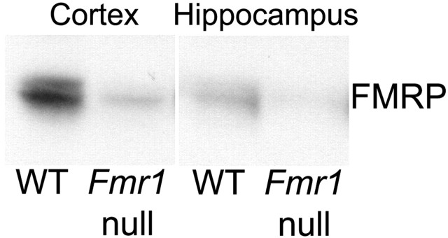
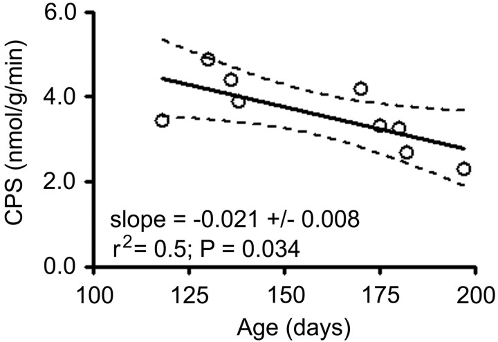
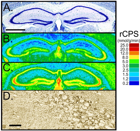
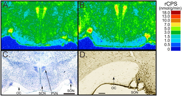
Methylation-induced transcriptional silencing of the fragile X mental retardation-1 (Fmr1) gene leads to absence of the gene product, fragile X mental retardation protein (FMRP), and consequently fragile X syndrome (FrX), an X-linked inherited form of mental retardation. Absence of FMRP in Fmr1 null mice imparts some characteristics of the FrX phenotype, but the precise role of FMRP in neuronal function remains unknown. FMRP is an RNA-binding protein that has been shown to suppress translation of certain mRNAs in vitro. We applied the quantitative autoradiographic L-[1-14C]leucine method to the in vivo determination of regional rates of cerebral protein synthesis (rCPS) in adult wild-type (WT) and Fmr1 null mice at 4 and 6 months of age. Our results show a substantial decrease in rCPS in all brain regions examined between the ages of 4 and 6 months in both WT and Fmr1 null mice. Superimposed on the age-dependent decline in rCPS, we demonstrate a regionally selective elevation in rCPS in Fmr1 null mice. Our results suggest that the process of synaptic pruning during young adulthood may be reflected in decreased rCPS. Our findings support the hypothesis that FMRP is a suppressor of translation in brain in vivo.


Figures

Figure 1.
Figure 1.
Western blots of protein extracts from cortex and hippocampus of WT and Fmr1 null mice demonstrating the specificity of the FMRP antibody.
Figure 2.
Figure 2.
Typical steady-state experiments for the determination of λ in WT (A) and Fmr1 null (B) mice illustrating the achievement and maintenance of a constant SA of [3H]leucine (□) over 120 min and the relatively constant arterial plasma leucine concentration (▵) over this interval. The figure also shows the SA of the tRNA-bound leucine (•) in brain at the end of the experiment. In both genotypes, the tRNA-bound leucine SA is considerably below that of the arterial plasma, indicating that there is significant recycling of unlabeled leucine derived from protein breakdown back into the precursor amino acid pool.
Figure 3.
Figure 3.
Correlation of CPS with age in young adult WT mice. Each point represents a measured value of global CPS in a single animal. The solid line is the best-fitting regression line. Dashed lines are the 95% confidence limits.
Figure 4.
Figure 4.
Digitized autoradiograms of coronal sections at the level of the dorsal hippocampus from representative 6-month-old WT (B) and Fmr1 null (C) mice. Images have been color coded for rCPS. The color bar (right) provides the calibration scale for the range of values of rCPS in nanomoles per gram per minute for each color. For comparison with the distribution of FMRP in a WT mouse brain, we used immunohistochemistry with a monoclonal antibody to FMRP. Immunostaining of FMRP in the CA1 sector of the pyramidal cell layer is shown in D. The CA1, CA2, and CA3 sectors of the pyramidal cell layer can be located in a thionin-stained section from a WT mouse (A). Scale bars: (in A) A-C, 1.0 mm; D, 35 μm. In A-D, dorsal is up and the left side of the brain is on the left.
Figure 5.
Figure 5.
Digitized autoradiograms of coronal sections at the level of the anterior hypothalamus from representative 6-month-old WT (A) and Fmr1 null (B) mice. Images have been color coded for rCPS. The color bar (right) provides the calibration scale for the range of rCPS in nanomoles per gram per minute for each color. For comparison with the distribution of FMRP in a WT mouse brain, we used immunohistochemistry with a monoclonal antibody to FMRP (D). Supraoptic (SON), paraventricular (PVN), and suprachiasmatic (SCN) nuclei and optic chiasm (OC) can be located in a thionin-stained section from a WT mouse (C). Scale bars: (in C) A-C, 0.5 mm; D, 75 μm. In A-D, dorsal is up and the left side of the brain is on the left.

References

    1. Agranoff BW (1981) Learning and memory: biochemical approaches. In: Basic neurochemistry (Seigel GJ, Albers RW, Agranoff BW, Katzman R, eds), pp 801-820. Boston: Little, Brown and Company.
    1. Antar LN, Afroz R, Dictenberg JB, Carroll RC, Bassell GJ (2004) Metabotropic glutamate receptor activation regulates fragile X mental retardation protein and frma mRNA localization differentially in dendrites and at synapses. J Neurosci 24: 2648-2655. - PMC - PubMed
    1. Aschrafi A, Cunningham BA, Edelman GM, Vanderklish PW (2005) The fragile X mental retardation protein and group 1 metabotropic glutamate receptors regulate levels of mRNA granules in brain. Proc Natl Acad Sci USA 102: 2180-2185. - PMC - PubMed
    1. Baubet V, Grange E, Sermet E, Giaume M, Gay N, Bobillier P (1996) Wide-spread increase in brain protein synthesis following acute immobilization stress in adult rat brain. Neurosci Lett 219: 187-190. - PubMed
    1. Bear M, Huber KM, Warren ST (2004) The mGluR theory of fragile X mental retardation. Trends Neurosci 27: 370-377. - PubMed

Publication types