. 2005 Jul;16(7):3438-53.
doi: 10.1091/mbc.e04-10-0894. Epub 2005 May 18.

Atg17 regulates the magnitude of the autophagic response

Affiliations

Atg17 regulates the magnitude of the autophagic response

Heesun Cheong et al. Mol Biol Cell. 2005 Jul.

Abstract

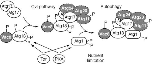
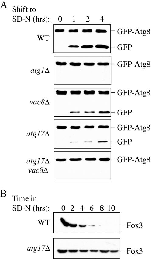
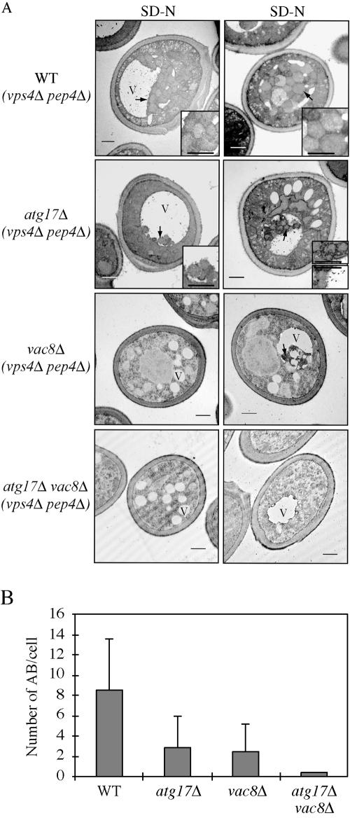
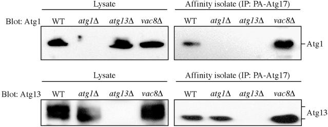
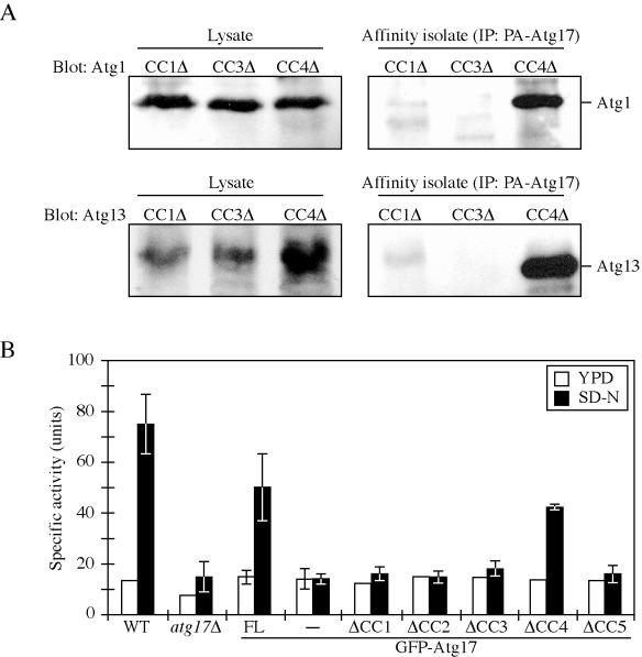
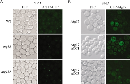
Autophagy is a catabolic process used by eukaryotic cells for the degradation and recycling of cytosolic proteins and excess or defective organelles. In yeast, autophagy is primarily a response to nutrient limitation, whereas in higher eukaryotes it also plays a role in developmental processes. Due to its essentially unlimited degradative capacity, it is critical that regulatory mechanisms are in place to modulate the timing and magnitude of the autophagic response. One set of proteins that seems to function in this regard includes a complex that contains the Atg1 kinase. Aside from Atg1, the proteins in this complex participate primarily in either nonspecific autophagy or specific types of autophagy, including the cytoplasm to vacuole targeting pathway, which operates under vegetative growth conditions, and peroxisome degradation. Accordingly, these proteins are prime candidates for factors that regulate the conversion between these pathways, including the change in size of the sequestering vesicle, the most obvious morphological difference. The atg17delta mutant forms a reduced number of small autophagosomes. As a result, it is defective in peroxisome degradation and is partially defective for autophagy. Atg17 interacts with both Atg1 and Atg13, via two coiled-coil domains, and these interactions facilitate its inclusion in the Atg1 complex.


Figures

Figure 1.
Figure 1.
Schematic model depicting assembly of the Atg1 kinase complex. Atg17 first interacts with the Atg13-Vac8 proteins. Atg13 and Atg1 may be phosphorylated by Tor and/or PKA (Budovskaya et al., 2004; Schmelzle et al., 2004) or by downstream effectors. The highly phosphorylated form of Atg13 has a lower affinity for Atg1 (Kamada et al., 2000) and this form of the complex may promote the Cvt pathway. Partial dephosphorylation of Atg13 and Atg1 allows the assembly of the putative holo-complex that triggers autophagy. Atg17 is depicted as interacting with Atg1 via Atg13, but the interaction may be direct (see text). Factors that have previously been characterized as being relatively specific for the Cvt pathway are depicted in gray. The timing of assembly of the Atg11, Atg20, and Atg24 proteins is not known as denoted by dotted lines. See text for additional details.
Figure 2.
Figure 2.
The atg17Δ mutant is partially defective in autophagy. (A) The atg17Δ mutant is not defective in the Cvt pathway. Wild-type (SEY6210), atg1Δ (WHY001), vac8Δ (YTS178), atg17Δ (CWY239), atg17Δ vac8Δ (CWY332), and atg17Δ atg20Δ (CWY333) strains were grown in SMD and shifted to SD-N for the indicated times to induce autophagy. atg17Δ cells were not defective for processing of prApe1 by the Cvt or autophagy pathways. The atg17Δ mutation in combination with vac8Δ, but not atg20Δ, was blocked in the autophagic delivery of prApe1. (B) Pho8Δ60, a marker for nonspecific autophagy, indicates an autophagy defect in the atg17Δ strain. The wild-type (TN124), atg1Δ (HAY572), atg20Δ (D3Y112), vac8Δ (CWY278), atg17Δ (CWY279), and atg17Δ vac8Δ (HCY43) strains were grown in YPD and shifted to SD-N for 4 h. Samples were collected and protein extracts assayed for ALP activity. The results represent the mean and SD of three experiments.
Figure 3.
Figure 3.
Autophagy and pexophagy occur at a reduced level in atg17Δ mutant cells. (A) Wild-type (SEY6210), atg1Δ (WHY001), vac8Δ (YTS178), atg17Δ (CWY239), and atg17Δ vac8Δ (CWY332) strains expressing GFP-Atg8 from pGFP-Aut7(414) were grown in SMD lacking auxotrophic amino acids and shifted to SD-N medium. At the indicated times, aliquots were removed, the proteins precipitated with TCA, and resolved by SDS-PAGE. Full-length GFP-Atg8 and free GFP were detected by immunoblot by using anti-GFP antibodies as described in Materials and Methods. The band running just below full-length GFP-Atg8 is a cross-contaminant. (B) atg17Δ cells are deficient in peroxisome degradation. Wild-type (BY4742) and atg17Δ cells were shifted from oleic acid-containing medium to SD-N, and pexophagy was monitored by Western blot by using an antibody against peroxisomal thiolase (Fox3) as described in Materials and Methods.
Figure 4.
Figure 4.
(A) The atg17Δ mutant generates fewer and smaller autophagosomes. The wild-type (FRY143; vps4Δ pep4Δ), atg17Δ (HCY31; vps4Δ pep4Δ), vac8Δ (HCY35; vps4Δ pep4Δ), and atg17Δ vac8Δ (HCY36; vps4Δ pep4Δ) strains were grown to mid-log stage in YPD and transferred to SD-N medium for 4 h. Cells were fixed with permanganate and examined by electron microscopy as described in Materials and Methods. Arrows mark the locations of autophagic bodies. The bars in the main images and insets (2× magnification) represent 0.5 μm. V, vacuole. (B) Quantification of autophagic body accumulation. Fifty sections for each strain were scored for autophagic body (AB) accumulation.
Figure 5.
Figure 5.
Yeast two-hybrid analysis among Atg1 complex components. The wild-type (PJ69–4A), atg1Δ (DKY6901), atg13Δ (CWY277), atg17Δ (CWY270), or the vac8Δ (CWY276) strains were transformed with the empty AD or BD vectors or with the vectors containing full-length Atg1, Atg13, Atg17, or Vac8 as indicated. After selection on plates lacking uracil and leucine, the transformants were patched onto plates additionally lacking either adenine, or histidine and containing 10 mM 3-aminotriazole. The plates were grown for 5 d at 30°C.
Figure 6.
Figure 6.
Atg17 interacts with Atg13 and Atg1. The wild-type (SEY6210), atg1Δ (WHY001), atg13Δ (CWY233), and vac8Δ (YTS178) strains expressing protein A-tagged Atg17 or the empty vector were grown in SMD plus casamino acids (0.5%). Protein extracts (lysate) were prepared and incubated with IgG-Sepharose beads as described in MATERIALS AND METHODS. The resulting immunocomplexes (affinity isolate) were resolved by SDS-PAGE and analyzed by Western blot by using serum directed against Atg1 or Atg13. No bands were detected from samples prepared from the strains carrying the empty protein A vector.
Figure 7.
Figure 7.
Regions in the coiled-coil domains of Atg17 are needed for interaction with its binding partners. (A) Schematic representation of Atg13, including the location of domains that bind Atg1, Atg17, and Vac8. Regions of Atg13 present in the activation domain (AD) two-hybrid protein constructs are depicted. The ability of the corresponding proteins to interact with the full-length Atg17 and Vac8 or an N-terminally truncated Atg1 binding domain two-hybrid protein is indicated on the right. The data for interactions between Atg1 and Atg13 are from Kamada et al. (2000) and for Atg13 and Vac8 are from Scott et al. (2000). ND, not determined. (B) A schematic representation of Atg17, indicating the location of predicted coiled-coil domains, is shown at the top. Regions of Atg17 deleted from the binding domain (BD) two-hybrid protein construct are depicted. The ability of the corresponding proteins to interact with the full-length Atg1 or Atg13 activation domain two-hybrid protein is indicated on the right. “+” means that the strain containing the two plasmids was able to grow on plates lacking histidine, and “–” indicates an inability to grow after 5 d. A deletion of the first or third coiled-coil domain in Atg17 blocked interaction with both Atg13 and Atg1.
Figure 8.
Figure 8.
The first and third Atg17 coiled-coil domains are required for interaction with Atg1 and Atg13 and for autophagic function. (A) Affinity isolation analysis. The atg17Δ (CWY239) strains expressing the indicated protein A-tagged coiled-coil domain-deleted Atg17 proteins were grown in SMD lacking auxotrophic amino acids. Protein extracts (lysate) were prepared and incubated with IgG-Sepharose beads as described in Materials and Methods. The resulting immunocomplexes (affinity isolate) were resolved by SDS-PAGE and analyzed by Western blot by using serum directed against Atg1 or Atg13. (B) Pho8Δ60 assay. The wild-type (TN124) and atg17Δ (CWY279) strain or the atg17Δ strain expressing full-length Atg17 or coiled-coil domain-deleted Atg17 in pRS426-CuGFP plasmids were grown in SMD lacking auxotrophic amino acids and shifted to SD-N for 4 h. The atg17Δ strain transformed with the pRS426-CuGFP empty plasmid was used as a negative control. Samples were collected and protein extracts assayed for ALP activity as described in Materials and Methods. The results represent the mean and SD of three experiments.
Figure 9.
Figure 9.
Atg17 localization depends on the first coiled-coil domain. Cells from (A) wild-type (CWY241), atg1Δ (CWY242), and atg13Δ (CWY263) strains were grown in YPD. The cells were examined by fluorescence microscopy as described in Materials and Methods. (B) For localization of Atg17 mutants, the atg17Δ (CWY239) strain expressing full length Atg17 or coiled-coil domain deleted Atg17 on pRS416-CuGFP plasmids was grown in SMD lacking auxotrophic amino acids and examined as described in A. DIC, differential interference contrast.
Figure 10.
Figure 10.
Localization of Atg1 and Atg9 in the atg17Δ mutant. (A) Wild-type (PSY143), atg17Δ (HCY032), atg13Δ (HCY040), and vac8Δ (HCY042) cells expressing Atg1-GFP were grown in YPD and shifted to SD-N for 2 h before microscopy. Atg1 showed both a punctate and diffuse cytosolic localization. The data for localization of Atg1-GFP in atg13Δ or vac8Δ were essentially the same as those shown for wild-type and atg17Δ. (B) Atg9 cycling is defective in cells lacking Atg17. Integrated Atg9-YFP was expressed in wild-type (FRY136), atg1Δ (FRY138), atg17Δ (KTY89), and atg1Δ atg17Δ (JLY4) cells. The cells were grown in YPD or appropriate selective media to mid-log phase and FM 4-64 was added to the culture medium for 15 min, after which the cells were pelleted and resuspended in YPD or SMD, and further incubated for 30 min, followed by imaging by fluorescence microscopy. In the merged panels Atg9-YFP is shown in green to facilitate visualization relative to the FM 4-64 dye. DIC, differential interference contrast.

References

    1. Abeliovich, H., Dunn, W. A., Jr., Kim, J., and Klionsky, D. J. (2000). Dissection of autophagosome biogenesis into distinct nucleation and expansion steps. J. Cell Biol. 151, 1025–1034. - PMC - PubMed
    1. Abeliovich, H., Zhang, C., Dunn, W. A., Jr., Shokat, K. M., and Klionsky, D. J. (2003). Chemical genetic analysis of Apg1 reveals a non-kinase role in the induction of autophagy. Mol. Biol. Cell 14, 477–490. - PMC - PubMed
    1. Baba, M., Osumi, M., Scott, S. V., Klionsky, D. J., and Ohsumi, Y. (1997). Two distinct pathways for targeting proteins from the cytoplasm to the vacuole/lysosome. J. Cell Biol. 139, 1687–1695. - PMC - PubMed
    1. Baba, M., Takeshige, K., Baba, N., and Ohsumi, Y. (1994). Ultrastructural analysis of the autophagic process in yeast: detection of autophagosomes and their characterization. J. Cell Biol. 124, 903–913. - PMC - PubMed
    1. Babst, M., Wendland, B., Estepa, E. J., and Emr, S. D. (1998). The Vps4p AAA ATPase regulates membrane association of a Vps protein complex required for normal endosome function. EMBO J. 17, 2982–2993. - PMC - PubMed

Publication types

MeSH terms