Comparative Study
. 2005 May 31;102(22):8030-5.
doi: 10.1073/pnas.0503021102. Epub 2005 May 18.

Carbonic anhydrase XIV is enriched in specific membrane domains of retinal pigment epithelium, Muller cells, and astrocytes

Affiliations
Comparative Study

Carbonic anhydrase XIV is enriched in specific membrane domains of retinal pigment epithelium, Muller cells, and astrocytes

Erlend A Nagelhus et al. Proc Natl Acad Sci U S A. .

Abstract

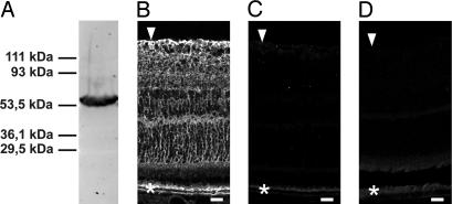
Carbonic anhydrases (CAs) are ubiquitous enzymes important to many cell types throughout the body. They help determine levels of H(+) and HCO(-)(3) and thereby regulate intracellular and extracellular pH and volume. CA XIV, an extracellular membrane-bound CA, was recently shown to be present in brain and retina. Here, we analyze the subcellular distribution of CA XIV in retina by high-resolution immunogold cytochemistry and show that the distribution in retina (on glial cells but not neurons) is different from that reported for brain (on neurons but not glia). In addition, CA XIV is strongly expressed on retinal pigment epithelium (RPE). The specific membrane domains that express CA XIV were endfoot and nonendfoot membranes on Muller cells and astrocytes and apical and basolateral membranes of RPE. Gold particle density was highest on microvilli plasma membranes of RPE, where it was twice that of glial endfoot and Muller microvilli membranes and four times that of other glial membrane domains. Neither neurons nor capillary endothelial cells showed detectable labeling for CA XIV. This enrichment of CA XIV on specific membrane domains of glial cells and RPE suggests specialization for buffering pH and volume in retinal neurons and their surrounding extracellular spaces. We suggest that CA XIV is the target of CA inhibitors that enhance subretinal fluid absorption in macular edema. In addition, CA XIV may facilitate CO(2) removal from neural retina and modulate photoreceptor function.


Figures

Fig. 1.
Fig. 1.
Selectivity of antibody to CA XIV. (A) Immunoblot of membrane fraction of mouse retina reveals a single band at ≈54 kDa. (B) Immunofluorescence of CA XIV in mouse retina. (C and D) No specific labeling remains after preabsorption of the primary antibody with the immunizing recombinant CA XIV (C) or after omission of the primary antibody (D). Weak autofluorescence occurs in the RPE (C and D). Arrowheads indicate inner limiting membrane, and asterisks indicate RPE. (Scale bars: 20 μm.)
Fig. 2.
Fig. 2.
Distribution of CA XIV immunoreactivity in the retina. The same antibody as in Fig. 1 is shown. (A) Immunolabeling extends throughout the neural retina and RPE (*). Note the dense labeling of RPE, outer plexiform layer, and inner limiting membrane (arrowheads). The immunofluorescent signal is particularly intense at the apical membrane of the RPE and is also quite pronounced at the inner aspect of the subretinal space [corresponding to the microvilli of the Müller cells (double arrowhead)]. Arrow indicates large vessel. (B) Interference optics, the same section as in A (arrows in A and B denote the same vessel). GCL, ganglion cell layer; IPL, inner plexiform layer; INL, inner nuclear layer; OPL, outer plexiform layer; ONL, outer nuclear layer; PhL, photoreceptor layer. (C) CA XIV immunolabeling in the outer retina. Note tiny immunopositive processes (double arrowhead) delimiting the inner aspect of the subretinal space. No labeling is seen over photoreceptor outer segments in PhL. The RPE (*) is intensely labeled apically and moderately labeled basally. (D) Interference optics, the same section as in C. OLM, outer limiting membrane. (Scale bars: A,50 μm; C,10 μm.)
Fig. 3.
Fig. 3.
Electron micrographs showing CA XIV immunoreactivity in the retina. (A and B) Gold particles signaling CA XIV are associated with subvitreal endfeet membranes of astrocytes (As) and Müller cells (Mü). Arrowheads indicate vitreal surface. (C and D) CA XIV immunopositive Müller processes in the outer plexiform layer are sandwiched between immunonegative photoreceptor terminals (Ph). (E) CA XIV is expressed in perivascular endfeet of Müller cells, with a stronger labeling in the membrane facing the vessel than in the membrane facing the neuropil. Double arrow spans the distance between the two membrane domains. Endothelial cells (End) and pericytes (Pe) are immunonegative. (Scale bars: 0.25 μm.)
Fig. 4.
Fig. 4.
CA XIV immunogold labeling in the outer retina. (A and B) Gold particles decorate microvilli (*) of Müller cells. Photoreceptor inner segments (IS) are immunonegative. (C and D) Microvilli (arrow in C) of RPE cells display stronger immunogold signals than the basolateral epithelial membrane (crossed arrow in D). Photoreceptor outer segments (OS) do not show immunogold labeling significantly above background level (compare Fig. 5B). Dark bodies represent pigment granules. (Scale bars: A and B, 0.25 μm; C and D, 0.5 μm.)
Fig. 5.
Fig. 5.
Quantitative analysis of CA XIV immunogold labeling in specific membrane domains. Values along the abscissa represent the mean number of gold particles per micrometer of membrane, recorded at a primary magnification of ×34,500. Number of profiles and SEM are indicated. AsEf, membranes of astrocyte endfeet facing vitreal surface; As, lateral membranes of astrocyte endfeet; EndLu, luminal membranes of endothelial cells; EndAb, abluminal membranes of endothelial cells; MüEf, membranes of Müller cell endfeet facing vitreal surface; MüGcl, lateral membranes of Müller cell endfeet; MüInl, Müller cell membranes in the inner nuclear layer; MüPvEf, perivascular membranes of Müller cell endfeet; MüPv, lateral membranes of perivascular Müller cell endfeet; MüOpl, Müller cell membranes in the outer plexiform layer; MüMv, membranes of Müller cell microvilli; PhIs, membranes of photoreceptor inner segments; RPEa, apical membranes of RPE; RPEb, basolateral membranes of RPE. The statistical analysis of these data is presented in Fig. 7.
Fig. 6.
Fig. 6.
Distribution of CA XIV signaling gold particles. Shown is particle distribution along an axis perpendicular to the perivascular plasma membrane of Müller cell endfeet (MüPvEf; A) and the plasma membrane of photoreceptor inner segments (PhIs; B) (compare Fig. 5). The ordinate indicates number of gold particles per bin (bin width, 4 nm; cytoplasmic side negative). The gold particle density drops to background level ≈25 nm from the midpoint of the plasma membrane [corresponding to the size of the antibody bridge between the epitope and the corresponding gold particle (24)]. The level of background labeling is slightly higher over cytoplasmic matrix (left in A) than over the perivascular basal lamina (right in A). There is no detectable peak over the photoreceptor plasma membrane (B).

Similar articles

Cited by

References

    1. Chesler, M. (2003) Physiol. Rev. 83, 1183–1221. - PubMed
    1. Wadiche, J. I., Amara, S. G. & Kavanaugh, M. P. (1995) Neuron 15, 721–728. - PubMed
    1. Magleby, K. L. (2004) Trends Neurosci. 27, 231–233. - PubMed
    1. Svichar, N. & Chesler, M. (2003) Glia 41, 415–419. - PubMed
    1. McMurtrie, H. L., Cleary, H. J., Alvarez, B. V., Loiselle, F. B., Sterling, D., Morgan, P. E., Johnson, D. E. & Casey, J. R. (2004) J. Enzyme Inhib. Med. Chem. 19, 231–236. - PubMed

Publication types

LinkOut - more resources