. 2005 May 19;435(7040):360-4.
doi: 10.1038/nature03595.

The Ter mutation in the dead end gene causes germ cell loss and testicular germ cell tumours

Affiliations

The Ter mutation in the dead end gene causes germ cell loss and testicular germ cell tumours

Kirsten K Youngren et al. Nature. .

Abstract

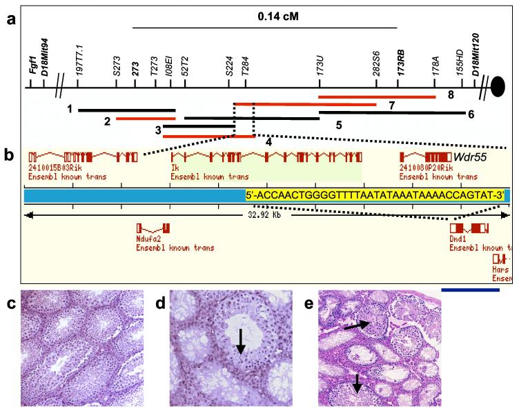
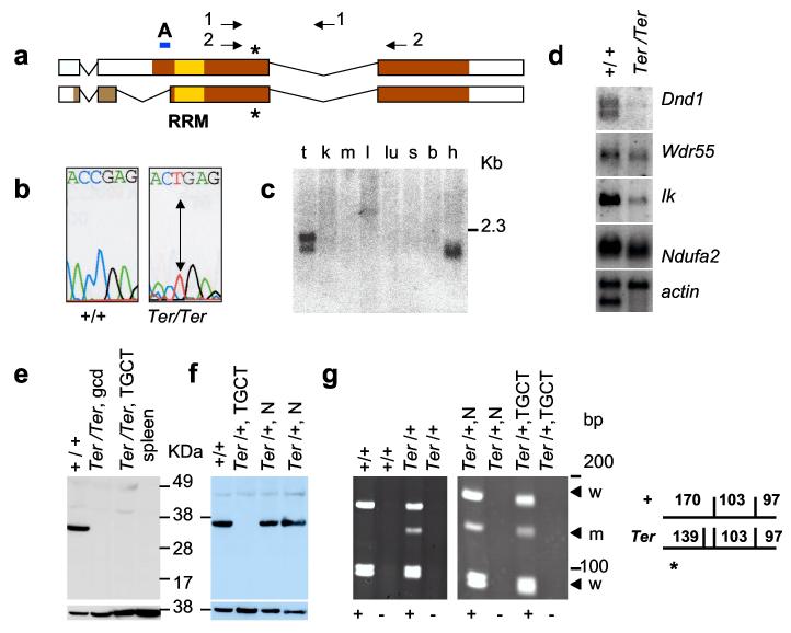
In mice, the Ter mutation causes primordial germ cell (PGC) loss in all genetic backgrounds. Ter is also a potent modifier of spontaneous testicular germ cell tumour (TGCT) susceptibility in the 129 family of inbred strains, and markedly increases TGCT incidence in 129-Ter/Ter males. In 129-Ter/Ter mice, some of the remaining PGCs transform into undifferentiated pluripotent embryonal carcinoma cells, and after birth differentiate into various cells and tissues that compose TGCTs. Here, we report the positional cloning of Ter, revealing a point mutation that introduces a termination codon in the mouse orthologue (Dnd1) of the zebrafish dead end (dnd) gene. PGC deficiency is corrected both with bacterial artificial chromosomes that contain Dnd1 and with a Dnd1-encoding transgene. Dnd1 is expressed in fetal gonads during the critical period when TGCTs originate. DND1 has an RNA recognition motif and is most similar to the apobec complementation factor, a component of the cytidine to uridine RNA-editing complex. These results suggest that Ter may adversely affect essential aspects of RNA biology during PGC development. DND1 is the first protein known to have an RNA recognition motif directly implicated as a heritable cause of spontaneous tumorigenesis. TGCT development in the 129-Ter mouse strain models paediatric TGCT in humans. This work will have important implications for our understanding of the genetic control of TGCT pathogenesis and PGC biology.


Figures

Figure 1.
Figure 1.
Gonadal phenotypes of Ter/Ter males. (a) Testis of 4-week old Ter/Ter male showing lack of germ cells (arrow points to Sertoli cell only phenotype) in the seminiferous tubules. (b,c) Histological sections through testicular germ cell tumours (TGCTs) from 4-week old 129-Ter/Ter male mice. Tissue types observed includes cartilage (arrow in b) and neuroepithelia (* in c). Arrow in (c) indicates germ cell deficient seminiferous tubules adjacent to neuroepithelial cells of the tumour. (d) GFP-tagged PGCs. Genital ridges of E12.5 embryos showing GFP expression in PGCs of 129-+/+;Oct4-GFP male, (e) 129-Ter/Ter;Oct4-GFP male, and (f) 129-Ter/Ter;Oct4-GFP female. The arrow indicates one of the genital ridges of the dissected embryo.
Figure 2.
Figure 2.
Positional cloning of Ter. (a) Physical map of the Ter locus. The 129-derived (RG-MBAC) BAC contig: 1 = 197J23, 2 = 173P21, 3 = 224K2, 4 = 284F9, 5 = 52N10, 6 = 368A6, 7 = 282N17, 8 = 273D11. (b) Ensembl gene map of the overlapping region of BACs 284F9 and 282N17. Overlapping bases of the last exons of Wdr55 and Dnd1 are marked in yellow. Blue line indicates the DNA fragment used in Tg(Dnd1)1Matn. (c) Testes histology of Ter/Ter with BAC 284F9 and sibling (d) also with transgene where rescue occurs in a subset of seminiferous tubules (arrow). (e) Histology of Ter/Ter testes with Tg(Dnd1)1Matn (arrow showing normal seminiferous tubule).
Figure 3.
Figure 3.
Expression of Dnd1 in normal tissues and TGCTs. (a) Comparison of DND1 isoforms with accession numbers BC034897 (top) and AY321066 (bottom). Colored regions indicate protein coding regions; yellow box marks RRM. Asterisk indicates amino acids 178 and 190, of the two isoforms, that is mutated (R178X) in Ter. Arrows indicate primers 4.13a (1) and A25 (2). A indicates the region used as peptide epitope to generate anti-DND1 antibody. (b) C to T mutation in Ter/Ter introduces stop codon. (c) Mouse tissue Northern blot for Dnd1. mRNAs are from testes (t), kidney (k), muscle (m), liver (l), lung (lu), spleen (s), brain (b) and heart (h). (d) Northern blot of mRNA from 129-+/+ testes and 129-Ter/Ter tumours. (e) Western blotting with anti-DND1 antibody of tissue lysates from normal (+/+) testes of 129-+/+, germ cell deficient (gcd) testes of B6.129-Ter/Ter congenic strain, bilateral TGCTs from 129-Ter/Ter mice, and spleen of 129-+/+. Lower panel shows control for loading using anti-β-actin. (f) Western blot with anti-DND1 of normal testes of 129-+/+ (+/+), tumour in the testes of 129-Ter/+ (Ter/+, TGCT), the normal contralateral testes (Ter/+, N), and normal testes of another 129-Ter/+ mouse (Ter/+, N). Lower panel shows the same blot re-hybridized with mouse anti-β-actin. (g) (left) RT-PCR of RNA from normal testes of 129-+/+ and 129-Ter/+ mice using primers A25 (2 in Fig. 3a) followed by Dde1 digestion. This produces fragments of 170, 103 and 97 bp from the + allele and 139, 103, 97 and 31 bp (not seen on gel) from the Ter allele. For controls, PCR reactions were performed on samples which included (+) or did not include Superscript II (-) during RT, as indicated below the panels. (right) Total RNA from normal and TGCT-bearing testes of the same 129-Ter/+ mouse was amplified by RT-PCR followed by Dde1 digestion and electrophoresis. The arrows indicate the wild-type (w) and Ter (m) allele.
Figure 4.
Figure 4.
Expression of Dnd1 in embryonic gonads. (a) Whole-mount in situ hybridization of E11.5 XY and (b) E11.5 XX genital ridge (g) and mesonephros (m), in which Dnd1 expression is localized to the gonad. (c) Whole-mount in situ hybridization of an E12.5 XY gonad (t), in which Dnd1 is localized to testis cords (tc), whereas Dnd1 expression in (d) E12.5 XX gonad (o) has a broad distribution throughout the gonad. (e) Whole-mount in situ hybridization of an E14.5 XY gonad, in which Dnd1 is localized to testis cords, in contrast to (f) E14.5 XX gonads in which Dnd1 expression is reduced. (g) and (h) are section in situ hybridization of an E12.5 XY and E13.5 XY gonad respectively. Dnd1 is specific to developing testicular cords in the E12.5 and E13.5 XY gonads. Scale bars represent 500 μm.

References

    1. Sakurai T, Iguchi T, Moriwaki K, Noguchi M. The ter mutation first causes primordial germ cell deficiency in ter/ter mouse embryos at 8 days of gestation. Develop. Growth Differ. 1995;37:293–302. - PubMed
    1. Noguchi T, Noguchi M. A recessive mutation (ter) causing germ cell deficiency and a high incidence of congenital testicular teratomas in 129/Sv-ter mice. J. Natl. Cancer Inst. 1985;75:385–392. - PubMed
    1. Stevens LC. A new inbred subline of mice (129-terSv) with a high incidence of spontaneous congenital testicular teratomas. J. Natl. Cancer Inst. 1973;50:235–242. - PubMed
    1. Stevens LC, Mackensen JA. Genetic and environmental influences on teratocarcinogenesis in mice. J. Natl. Cancer Inst. 1961;27:443–453.
    1. Stevens LC. Origin of testicular teratomas from primordial germ cells in mice. J. Natl. Cancer Inst. 1967;38:549–552. - PubMed

Publication types

MeSH terms