. 2005 May 19;435(7040):374-9.
doi: 10.1038/nature03580.

Structure of the zinc-binding domain of an essential component of the hepatitis C virus replicase

Affiliations

Structure of the zinc-binding domain of an essential component of the hepatitis C virus replicase

Timothy L Tellinghuisen et al. Nature. .

Abstract

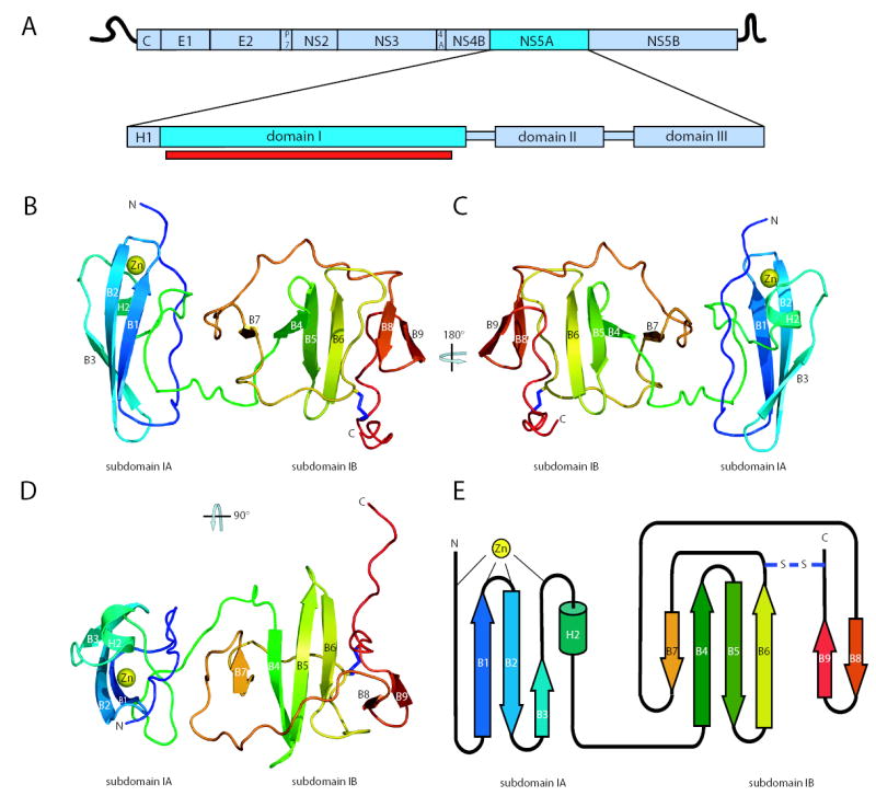
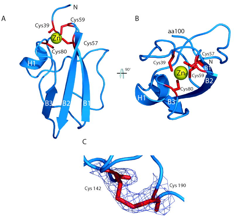
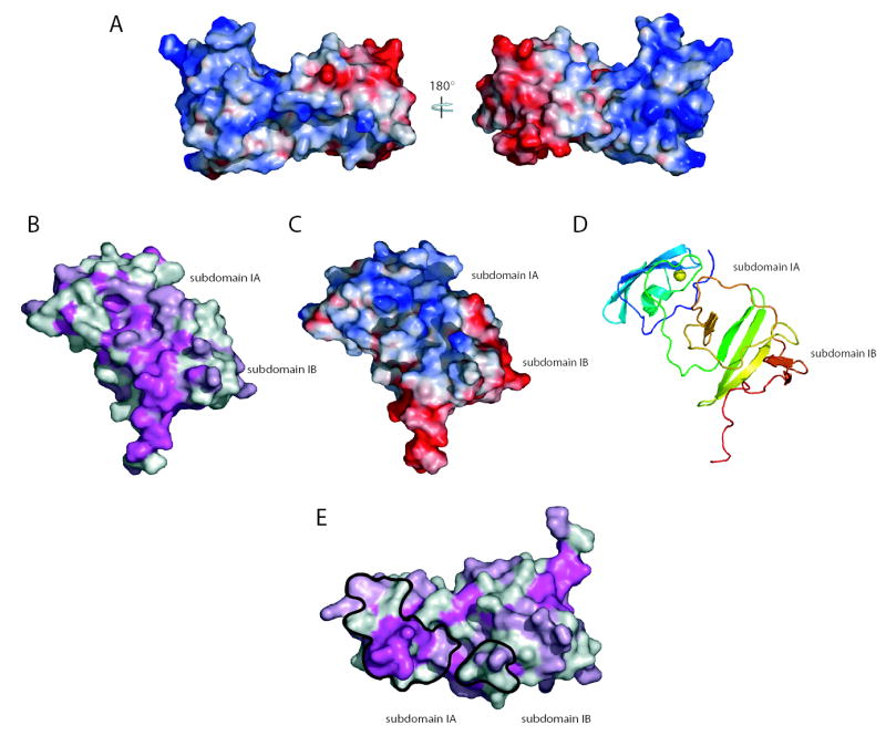
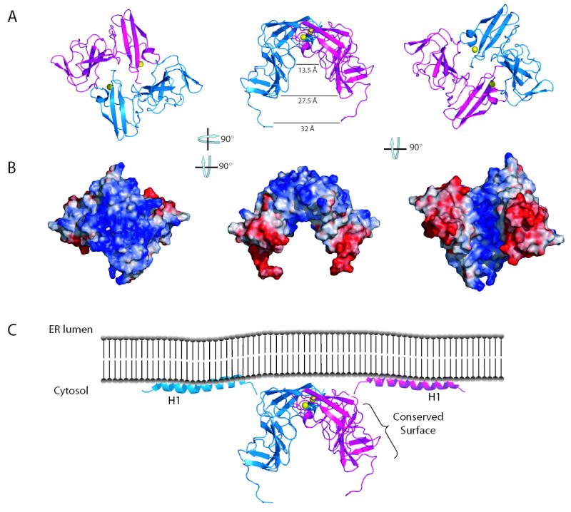
Hepatitis C virus (HCV) is a human pathogen affecting nearly 3% of the world's population. Chronic infections can lead to cirrhosis and liver cancer. The RNA replication machine of HCV is a multi-subunit membrane-associated complex. The non-structural protein NS5A is an active component of HCV replicase, as well as a pivotal regulator of replication and a modulator of cellular processes ranging from innate immunity to dysregulated cell growth. NS5A is a large phosphoprotein (56-58 kDa) with an amphipathic alpha-helix at its amino terminus that promotes membrane association. After this helix region, NS5A is organized into three domains. The N-terminal domain (domain I) coordinates a single zinc atom per protein molecule. Mutations disrupting either the membrane anchor or zinc binding of NS5A are lethal for RNA replication. However, probing the role of NS5A in replication has been hampered by a lack of structural information about this multifunctional protein. Here we report the structure of NS5A domain I at 2.5-A resolution, which contains a novel fold, a new zinc-coordination motif and a disulphide bond. We use molecular surface analysis to suggest the location of protein-, RNA- and membrane-interaction sites.


Conflict of interest statement

Competing interest statement The authors declare that they have no competing financial interests.

Figures

Figure 1
Figure 1
An overview of the NS5A domain I structure. a. Schematic of HCV genome organization and domain structure of the NS5A protein. The portion of domain I in this structure is indicated by the red bar. b. Ribbon diagram of the structure of domain I. The polypeptide chain is colored from N-terminus (blue) to C-terminus (red). The coordinated zinc atom is shown in yellow. The disulfide bond is shown in blue. c. A 180° rotation showing the ‘back’ of domain I. d. A 90° rotation showing the ‘top down’ view of domain I. e. Domain I topology organization model.
Figure 2
Figure 2
The NS5A zinc-binding motif and disulfide bond. a. A view of subdomain 1A (amino acids 36-100) highlighting the zinc-binding motif. The zinc atom (yellow) is coordinated by four cysteine residues (red). b. A ‘top down’ view of the zinc ion coordination site showing caging of the zinc atom. c. Overlay of experimental electron density map on the model of the disulfide bond at 1σ. Amino acids cysteine 142 and 190 are labeled.
Figure 3
Figure 3
Molecular surfaces of domain I. a. Domain I surface potential, acidic (red), neutral (white), and basic (blue) surfaces are shown. Two rotations of domain I are shown corresponding to the views in 1b and 1c. b. Surface of domain I colored by conservation; magenta ≥ 95% conservation, light pink = 75–95% conservation, and white ≤ 75% conservation. Domain I is oriented to show the most conserved molecular surface. c. A surface potential view of the image in b. d. A ribbon diagram view of the orientation in c and d. e. Conservation plot showing the dimer interface surfaces (black outline).
Figure 4
Figure 4
The NS5A domain I dimer reveals potential interaction surfaces. a. Ribbon diagrams of three rotations of the domain I dimer. b. Surface potential plots of the domain I dimer, views correspond to those show in a. Analysis of images in a and b reveal the dimer creates a relatively flat, basic surface near the N-terminus and a large groove between the two subdomain IB regions. c. Model of NS5A position relative to the ER membrane. The location of the conserved surface in figure 3 b is indicated. Helices are from the recent structure of the NS5A N-terminal helix.

References

    1. Anonymous World Health Organization - Hepatitis C: global prevalence. Wkly Epidemiol Rec. 1997;72:341–4. - PubMed
    1. Blight KJ, Kolykhalov AA, Rice CM. Efficient initiation of HCV RNA replication in cell culture. Science. 2000;290:1972–1974. - PubMed
    1. Lohmann V, et al. Replication of subgenomic hepatitis C virus RNAs in a hepatoma cell line. Science. 1999;285:110–113. - PubMed
    1. Lohmann V, Korner F, Dobierzewska A, Bartenschlager R. Mutations in hepatitis C virus RNAs conferring cell culture adaptation. J Virol. 2001;75:1437–1449. - PMC - PubMed
    1. Tellinghuisen TL, Rice CM. Interaction between hepatitis C virus proteins and host cell factors. Current Opinion in Microbiology. 2002;5:419–27. - PubMed

Publication types

MeSH terms

Associated data