. 2005 Jun;115(6):1522-31.
doi: 10.1172/JCI23769. Epub 2005 May 12.

Morphogenesis of the right ventricle requires myocardial expression of Gata4

Affiliations

Morphogenesis of the right ventricle requires myocardial expression of Gata4

Elisabeth M Zeisberg et al. J Clin Invest. 2005 Jun.

Abstract

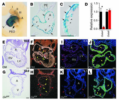
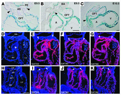
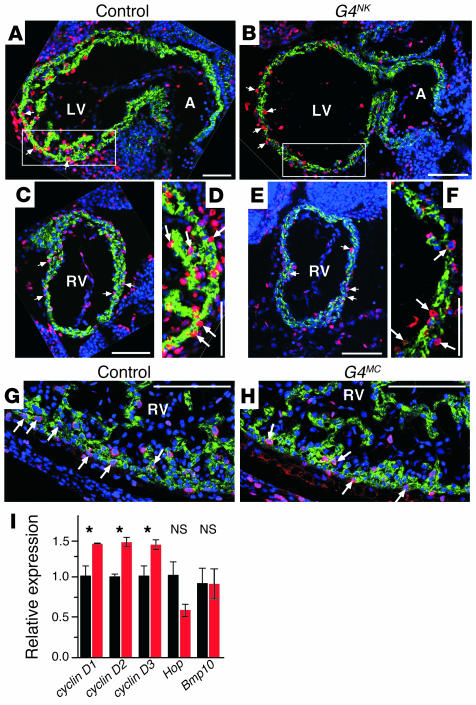
Mutations in developmental regulatory genes have been found to be responsible for some cases of congenital heart defects. One such regulatory gene is Gata4, a zinc finger transcription factor. In order to circumvent the early embryonic lethality of Gata4-null embryos and to investigate the role of myocardial Gata4 expression in cardiac development, we used Cre/loxP technology to conditionally delete Gata4 in the myocardium of mice at an early and a late time point in cardiac morphogenesis. Early deletion of Gata4 by Nkx2-5Cre resulted in hearts with striking myocardial thinning, absence of mesenchymal cells within the endocardial cushions, and selective hypoplasia of the RV. RV hypoplasia was associated with downregulation of Hand2, a transcription factor previously shown to regulate formation of the RV. Cardiomyocyte proliferation was reduced, with a greater degree of reduction in the RV than in the LV. Late deletion of Gata4 by Cre recombinase driven by the alpha myosin heavy chain promoter did not selectively affect RV development or generation of endocardial cushion mesenchyme but did result in marked myocardial thinning with decreased cardiomyocyte proliferation, as well as double-outlet RV. Our results demonstrate a general role of myocardial Gata4 in regulating cardiomyocyte proliferation and a specific, stage-dependent role in regulating the morphogenesis of the RV and the atrioventricular canal.


Figures

Figure 1
Figure 1
Early cardiomyocyte-restricted inactivation of Gata4 by Nkx2-5Cre. (AC) The spatial pattern of recombination driven by Nkx2-5Cre was determined using the R26RstoplacZ reporter. X-gal–stained E9.5 embryos were examined in whole-mount (A) or in transverse sections (B and C). Strong staining was detected in cardiomyocytes (arrows, B and C) of the atrium (A) and ventricle (V). Weaker activity was detected in the branchial arch (I–III), epithelium, and pharyngeal endoderm (PE). No Cre activity was detected in the proepicardial organ (PEO) or endocardium (arrowheads). (D) Efficient inactivation of Gata4 by Nkx2-5Cre. Gata4 and Gata6 expression was measured by qRT-PCR in RNA isolated from hearts of E9.5 control (Gata4flox/flox, black bars) or G4NK embryos (red bars). Gata4 expression was downregulated in G4NK embryos compared with that of controls (n = 3; *P < 0.05). (EH) In situ hybridization for Gata4. Gata4 expression was robust in myocardium (red arrowheads), endocardium (yellow arrowheads), and endocardial cushions (asterisk) of control embryos (E and F) but was not detectable in G4NK embryos (G and H). Shown are brightfield (E and G) and darkfield views (F and H). (IL) Immunofluorescent detection of Gata4 in control (I and J) and G4NK embryos (K and L). Gata4 staining was strongly reduced in cardiomyocytes (red arrowheads) and endocardial cells (yellow arrowheads) of G4NK embryos. J and L show the same field as I and K, with the addition of green labeling of cardiomyocytes (desmin). Red staining indicates Gata4; blue staining denotes nuclei; purple staining indicates colocalization of Gata4 in the nucleus. Original magnification, ×40 (A). Scale bars: 100 μm (B, C, and EL).
Figure 2
Figure 2
Phenotypic characterization of mutants after early cardiomyocyte-restricted Gata4 deletion. (A) Survival of mutant embryos after myocyte-restricted deletion of Gata4. Numbers indicate total number of embryos genotyped at each gestational age. Expected Mendelian incidence was 25%. (B and C) Unstained whole-mount control and G4NK embryos at E10.5, showing pericardial effusion (white arrows) of the mutant embryo. (DI) Control and mutant hearts, viewed from the left lateral (DF) and right anterior oblique (GI) positions. In control (G) and in 6 of 21 mutant (H) embryos systematically examined, the RV and OFT (arrows) connected normally to the right lateral aspect of the LV. In the remaining 15 of 21 mutant embryos, the RV was very hypoplastic or not apparent, and the OFT connected to the rostral aspect of the LV (I). The groove at the atrioventricular junction (arrowhead) was preserved in mutant embryos. (JP). H&GE-stained transverse sections at the level of the atrioventricular canal (J and K) and the OFT (NP). (L and M) Higher-magnification views of boxed regions in J and K, respectively. Mutant embryos had severe myocardial hypoplasia with reduced trabeculation. Arrowheads indicate the endocardial cushions, which were small and markedly hypocellular in mutant embryos (K and O) compared with those of controls (J and N). In some mutant embryos, the RV and OFT were thin walled but normally positioned (O), while in other mutant embryos the RV was not apparent, and the OFT arose in an abnormal position from the ventricular chamber (P). Embryos in DP had 31–32 somites. Original magnification, ×20 (B and C), ×60 (DI). Scale bars: 100 μm (JP).
Figure 3
Figure 3
Expression of Hand genes after early myocyte-restricted Gata4 deletion. (AF) Whole-mount (A and B) and section (CF) in situ hybridization for Hand2. In the hearts of control embryos (A, C, and E), Hand2 was predominantly expressed in the RV and OFT (arrows), although its expression was also detected in the LV. In G4NK embryos (B, D, and F), Hand2 expression was significantly decreased in the hypoplastic RV and OFT (arrows). Expression of Hand2 was detected in the branchial arches of both control and G4NK embryos (arrowheads). (GJ) Section in situ hybridization for Hand1. Hand1 expression was unchanged in mutant embryos compared with that in control embryos, localizing predominantly to the outer curvature of the LV (arrows), as well as to the branchial arches (arrowheads). Adjacent sections from the same mutant embryo were used in F and J. Brightfield (C, D, G, and H) and darkfield views (E, F, I, and J) are shown. Original magnification, ×32 (A and B). Scale bars: 100 μm (CJ).
Figure 4
Figure 4
Pattern of Gata4, Gata5, and Gata6 expression and Nkx2-5Cre activity in the secondary heart field. (AC) Nkx2-5Cre activation of the R26RstoplacZ reporter in the splanchnic mesoderm at its junction with the OFT (secondary heart field; arrowheads) at E9.5 (transverse section, A; sagittal section, B) and at E10.5 (sagittal section, C). Recombination in splanchnic mesoderm was detected in rare cells at E9.5 but was robust by E10.5. AS, aortic sac; BA, branchial arch. (DK) Adjacent transverse sections of the same E9.5 control embryo (22 somites) were hybridized to probes for Gata4, Gata5, and Gata6 (in situ signal displayed as red pseudocolor). Gata5 and Gata6 were detected in the splanchnic mesoderm (SM; arrowheads) and pharyngeal endoderm (data not shown). Gata4 was not expressed in the splanchnic mesoderm, but was detected in the myocardium and endocardium. Sections in DG were at the level of the caudal surface of the OFT (asterisk), while the plane of section in HK was slightly more cranial, sectioned at the junction of the OFT with the aortic sac. Scale bars: 100 μm.
Figure 5
Figure 5
Late cardiomyocyte-restricted Gata4 deletion by MHCαCre. (AD) Gata4 in situ hybridization (red pseudocolor) on sections from E12.5 G4MC and control embryos, showing inactivation of Gata4 in the myocardium after late cardiomyocyte-restricted Gata4 deletion (white arrowheads, compare A and C with B and D). Expression of Gata4 in endocardium (yellow arrowheads) and endocardial cushions (asterisks) was unchanged in G4MC hearts compared with controls. Red blood cells gave a nonspecific signal in darkfield (red arrowhead, B). Blue staining indicates nuclei. Blue arrowheads indicate epicardium. (E and F) Reduced myocardial expression of Gata4 in E12.5 G4MC embryos compared with littermate controls. In the control ventricle (E), Gata4 staining was detected in myocardium (white arrowheads), epicardium (blue arrowheads), and endocardium (yellow arrowheads). Some myocyte nuclei in control embryos appeared Gata4 negative (white arrow). In the mutant ventricle (F), Gata4 was present in epicardium and endocardium but not in myocardium. Red staining indicates Gata4; green staining indicates desmin; blue staining indicates nuclei (TOPRO3). (GL) Cardiac malformations after late cardiomyocyte-restricted Gata4 deletion. Transverse sections of E13.5 embryos were stained with H&GE. G4MC embryos (JL) had marked hypoplasia of the compact myocardium (arrowheads) and reduced trabecular myocardium compared with that of control embryos (GI). The aortic valve (AoV; H and K) and pulmonary valve (PV; I and L) arose from the RV in mutant embryos, which indicates the presence of a double-outlet RV. The atrioventricular canal was properly septated into right and left inflow channels by the endocardial cushions (asterisks), which were normally populated by mesenchymal cells (G and J). Scale bars: 100 μm (AD), 50 μm (E and F), 200 μm (GL).
Figure 6
Figure 6
Decreased myocyte proliferation in mutants lacking myocardial Gata4 expression. (AH) Cardiomyocyte staining was assessed by BrdU labeling (red stain). Nuclei were counterstained with TOPRO3 (blue stain), and cardiomyocytes were identified by desmin staining (green stain). (AF) Decreased cardiomyocyte proliferation in early myocyte-restricted Gata4-ablated (G4NK) hearts at E9.5 (control, 21.4 ± 1.2 somites; mutant, 20.8 ± 0.7 somites). Arrows indicate representative BrdU-positive myocyte nuclei. Sagittal sections from the same control (A, C, and D) or G4NK (B, E, and F) embryo are shown. (D and F) Higher magnification views of the boxed regions in A and B. (G and H) Decreased cardiomyocyte proliferation in late myocyte-restricted Gata4 ablated (G4MC) hearts at E12.5. Arrows indicate representative BrdU-positive myocyte nuclei. (I) Expression of candidate Gata4 target genes that control cardiomyocyte proliferation. RNA was isolated from E12.5 G4MC (red bars) and control hearts (black bars). *P < 0.05. Scale bars: 100 μm.
Figure 7
Figure 7
Gata4 regulation of cardiac gene expression. (A) Relative expression of transcription factors previously suggested to be Gata4 targets. RNA was isolated from embryo hearts at E9.5 (red bars, G4NK; black bars, Gata4flox/flox;Nkx2-5WT/WT controls) or at E12.5 (red bars, G4MC; black bars, Gata4flox/flox;MHCαCre controls). (B) Relative expression of structural genes putatively identified as Gata4 targets, in E12.5 hearts (red bars, G4MC; black bars, Gata4flox/flox;MHCαCre controls). Gene expression was measured by qRT-PCR and normalized to 18S ribosomal RNA. *P < 0.05. (C) Ectopic ANF expression in the OFT (blue arrow and shading), seen in 2 of 6 G4NK embryos examined at E9.5 by whole-mount in situ hybridization for ANF. Original magnification, ×32. (D) Model for the role of Gata4 and Nkx2-5 in the transcriptional regulation of ventricular formation. Distinct transcriptional programs control the formation of the LV and RV. Gata4 regulates RV morphogenesis, in part through Hand2, whereas Nkx2-5 regulates LV development in collaboration with Hand1. In addition to the specific role of Gata4 in RV formation, Gata4 regulates cardiomyocyte proliferation in both cardiac chambers via as-yet-unknown factors (X), which may be more dependent upon Gata4 in the RV than in the LV.

References

    1. Hoffman JI. Incidence of congenital heart disease: I. Postnatal incidence. Pediatr. Cardiol. 1995;16:103–113. - PubMed
    1. Waldo KL, et al. Conotruncal myocardium arises from a secondary heart field. Development. 2001;128:3179–3188. - PubMed
    1. Mjaatvedt CH, et al. The outflow tract of the heart is recruited from a novel heart-forming field. Dev. Biol. 2001;238:97–109. - PubMed
    1. Kelly RG, Brown NA, Buckingham ME. The arterial pole of the mouse heart forms from Fgf10-expressing cells in pharyngeal mesoderm. Dev. Cell. 2001;1:435–440. - PubMed
    1. Runyan RB, Markwald RR. Invasion of mesenchyme into three-dimensional collagen gels: a regional and temporal analysis of interaction in embryonic heart tissue. Dev. Biol. 1983;95:108–114. - PubMed

Publication types

MeSH terms