

Relationship between proliferative activity of cancer cells and clinicopathological factors in patients with esophageal squamous cell carcinoma
- PMID: 15902736
- PMCID: PMC4305667
- DOI: 10.3748/wjg.v11.i19.2956
Relationship between proliferative activity of cancer cells and clinicopathological factors in patients with esophageal squamous cell carcinoma
Abstract
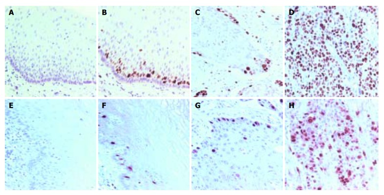
Aim: To assess whether the molecular markers of malignant tumors could improve the understanding of tumor characteristics, and to observe the characteristics of expression of cell cycle markers Ki-67 and cyclin A in esophageal carcinoma and to analyze the relationship between proliferative activity of cancer cells and clinicopathological factors.
Methods: Seventy of surgically resected esophageal squamous cell carcinoma (SCC) were examined by immunohistochemistry utilizing commercially available antibodies. Nuclear staining was regarded as a positive result. At least 50 fields in each tumor and non-tumor section were evaluated at a medium power (X200) to determine the proportion of tumor cells and the staining intensity of nuclei in the entire sections.
Results: Ki-67 and cyclin A were only expressed in base cells of normal esophageal mucosa. The positive immuno-staining of nuclei of SCC was significantly higher than that in normal esophageal mucosa (t=13.32 and t=7.52, respectively, P<0.01). The distribution of positively stained was more diffuse and stronger in poorly differentiated SCC. Both Ki-67 and cyclin A expressions were related to histological grades of tumors (t=3.5675 and t=3.916; t=2.13, respectively, P<0.05) but not to the sex and age of the patients, tumor size, lymphatic invasion, location, or stage grouping.
Conclusion: The proliferative activity of cancer cells may be understood by immunohistochemistry of Ki-67 and cyclin A in Chinese patients with esophageal SCC. These cell cycle markers may serve as an indicator of cancer cell proliferation rate. The overexpression of cell cycle markers Ki-67 and cyclin A suggests the poor SCC differentiation in patients with esophageal carcinoma.
Figures
References
-
- Tubiana M, Courdi A. Cell proliferation kinetics in human solid tumors: relation to probability of metastatic dissemination and long-term survival. Radiother Oncol. 1989;15:1–18. - PubMed
-
- Gerdes J, Lemke H, Baisch H, Wacker HH, Schwab U, Stein H. Cell cycle analysis of a cell proliferation-associated human nuclear antigen defined by the monoclonal antibody Ki-67. J Immunol. 1984;133:1710–1715. - PubMed
-
- Cattoretti G, Becker MH, Key G, Duchrow M, Schlüter C, Galle J, Gerdes J. Monoclonal antibodies against recombinant parts of the Ki-67 antigen (MIB 1 and MIB 3) detect proliferating cells in microwave-processed formalin-fixed paraffin sections. J Pathol. 1992;168:357–363. - PubMed
-
- Parkin DM, Läärä E, Muir CS. Estimates of the worldwide frequency of sixteen major cancers in 1980. Int J Cancer. 1988;41:184–197. - PubMed
Publication types
MeSH terms
Substances
LinkOut - more resources
Full Text Sources
Medical
Research Materials
