. 2005 May;96(5):265-73.
doi: 10.1111/j.1349-7006.2005.00043.x.

Overexpression of MUC13 is associated with intestinal-type gastric cancer

Affiliations

Overexpression of MUC13 is associated with intestinal-type gastric cancer

Takahiro Shimamura et al. Cancer Sci. 2005 May.

Abstract

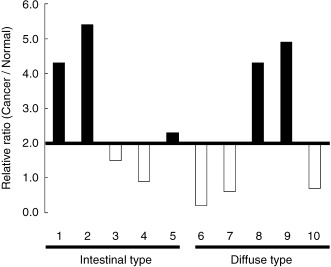
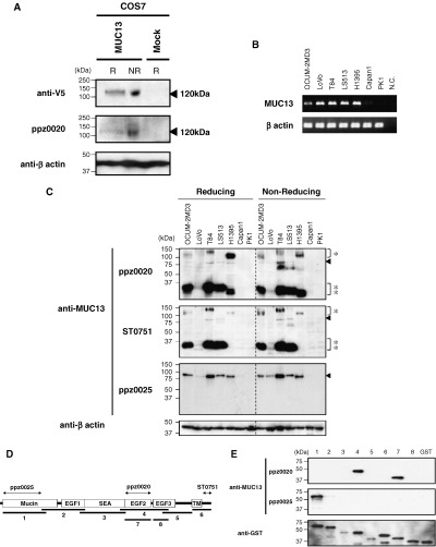
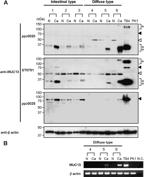
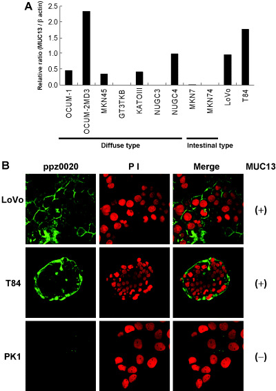
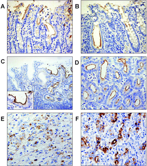
Mucins are secreted or transmembrane glycoproteins that are expressed mainly in the digestive tract. This family of proteins has been the focus of much gastric cancer research as some transmembrane mucins are implicated in tumorigenesis and make attractive targets for cancer diagnosis and therapeutics. Mucins have also been utilized to classify gastric cancer by differentiating between gastric and intestinal phenotypes. Here we show that transmembrane mucin MUC13 is upregulated in gastric cancer. By quantitative real-time reverse transcription-polymerase chain reaction and immunoblot analysis, overexpression of MUC13 was verified in more than half of the samples examined. In immunohistochemical analysis, MUC13 staining was observed in 74 of 114 cases of gastric cancer (64.9%), predominantly in intestinal type (P < 0.001), and in 9 of 10 cases of intestinal metaplasia, precancerous lesions of intestinal-type gastric cancer, but not observed in normal gastric mucosa. Moreover, MUC13 staining patterns characteristic of histological type were identified: staining was on the apical side of tubular glands in intestinal type and on the cytoplasm in diffuse type. These results suggest that MUC13 is a good differentiation marker for gastrointestinal mucosa and that it may have a causal role that correlates with two distinct gastric tumorigenesis pathways.


Figures

Figure 1
Figure 1
Overexpression of MUC13 in gastric cancer verified by quantitative reverse transcription‐polymerase chain reaction. Ten pairs of gastric cancer tissues and corresponding normal tissues were analyzed. Relative expression levels in cancer to normal tissue are indicated, and under 2.0 is denoted by blank and open bars, respectively. Note the expression of MUC13 was upregulated in five out of 10 pair samples, including both intestinal types and diffuse types.
Figure 2
Figure 2
Characterization of MUC13 protein with newly generated antibodies. (a) Immunoblot analysis of transiently expressed MUC13 protein. A full‐length cDNA of MUC13 and mock vector were transiently transfected into COS7 cells, and cell lysates were subjected to immunoblot analysis by anti‐V5 (upper panel), ppz0020 (middle panel) and anti‐β‐actin (lower panel). Note that the single band migrating at 120 kDa (arrow head) was detected only in MUC13‐transfected cells under both reducing (R) and non‐reducing (NR) conditions. (b) Reverse transcription‐polymerase chain reaction (RT–PCR) analysis of MUC13 in various cell lines. Coding full length MUC13 was amplified from the cDNA of seven cell lines (OCUM‐2 MD3, LoVo, T84, LS513, H1395, Capan1 and PK1). MUC13 expression was observed in five cell lines (OCUM‐2 MD3, LoVo, T84, LS513 and H1395), but not in two cell lines (Capan1 and PK1). β‐Actin was used as an internal control. (c) Detection of endogenous MUC13 protein with various antibodies. Endogenous MUC13 proteins in the seven cell lines above were examined with ppz0020 (upper panel), ST0751 (middle panel) and ppz0025 (lower panel) under R and NR conditions. Note ppz0020‐ and ST0751‐detected bands around 120 kDa (single asterisks), 80 kDa (arrowheads) and 35 kDa (double asterisks), while ppz0025 detected only an 80‐kDa band in MUC13‐expressing cells, as revealed by RT–PCR. β‐Actin was used as a loading control. (d) Schematic diagram of the putative domain structure of MUC13. MUC13 consists of a mucin domain (Mucin), three epidermal growth factor‐like domains (EGF 1–3), sea urchin sperm protein enterokinase agrin module (SEA) and transmembrane domain (TM). The epitope of each antibody as revealed by immunoblotting below is indicated by bipolar arrows. (e) Epitope mapping of anti‐MUC13 antibodies. Eight segments of MUC13 corresponding to the diagram above were fused with glutathione‐S‐transferase (GST) and blotted with ppz0020, ppz0025 and anti‐GST. β‐Actin was used as a loading control. N.C., negative control.
Figure 3
Figure 3
Expression of MUC13 protein in gastric cancer. (a) Detection of MUC13 protein in gastric cancer tissues. Immunoblot analysis was carried out on six pair samples of gastric cancer (Ca) and non‐cancerous (N) tissues. Two cancer cell lines, T84 and PK1, were utilized as positive and negative controls, respectively. Bands around 35 kDa (double asterisks) were detected more intensely in cancer, compared to normal, tissues in five of six pairs with ppz0020 (upper panel) and ST0751 (middle panel), and bands migrating at 80 kDa (closed arrowheads) were detected in cancer tissues in three of six pairs with ppz0025 (lower panel). 50‐kDa bands (open arrowheads) may represent cross‐reactivity associated with ppz0020 and ST0751 (see text). β‐Actin was included as a loading control. (b) Reverse transcription‐polymerase chain reaction analysis of MUC13 in gastric cancer. Three identical pairs (4–6) of gastric cancer tissues and corresponding normal tissues used in imunoblotting were analyzed. MUC13 expression was not observed in normal tissues, indicating that the 50‐kDa band detected above is due to cross‐reactivity of the antibodies. β‐Actin was included as an internal control. N.C., negative control.
Figure 4
Figure 4
Immunocytochemical analysis of MUC13. (a) Expression of MUC13 in gastric cancer cell lines. Seven cell lines derived from diffuse‐type gastric cancer (OCUM‐1, OCUM‐2 M, MKN45, GT3TKB, KATOIII, NUGC3 and NUGC4) and two derived from intestinal‐type gastric cancer (MKN7 and MKN74) were analyzed by quantitative reverse transcription‐polymerase chain reaction. Two colon cancer cell lines (LoVo and T84) were also analyzed as references. Relative expression levels of MUC13 to β‐actin are indicated. Note the expression of MUC13 was observed in five out of nine gastric cancer cell lines, relatively high in OCUM‐2 MD3 and NUGC4, and moderate in OCUM‐1, MKN45 and KATOIII. (b) Immunocytostaining analysis of endogenous MUC13. Subcellular localization of endogenous MUC13 was observed by confocal laser scan microscopy in MUC13‐positive (+) (LoVo, T84) and MUC13‐negative (–) (PK1) cell lines. Left panel, MUC13 staining using ppz0020; middle panel, nuclei stained with propidium iodide; right panel, merged image. Note endogenous MUC13 was detected only in MUC13‐expressing cells. MUC13 was localized to the plasma membrane of each cell in LoVo (upper panel), and to that of outer layer cells in T84 (middle panel). These photomicrographs depict ×63 magnification.
Figure 5
Figure 5
Immunohistochemical analysis of MUC13. (a–f) Immunohistochemical analysis of MUC13 in the gastrointestinal tract and in gastric cancer. Representative staining patterns are presented here. MUC13 staining was observed in the luminal surface of epithelial cells in the (a) small intestine, (b) large intestine and (c) in intestinal metaplasia of the stomach (right and inset) but not in the gastric foveolar epithelium (left). In gastric cancer, MUC13 staining was observed in (d) the apical surface of tubules of intestinal type, (e) in the cytoplasm of diffuse type and (f) in both the apical surface and cytoplasm of intestinal type with poor tubular gland formation. These photomicrographs depict (c) ×100 and (a,b,d,e) ×200 magnification.

References

    1. Parkin DM. Global cancer statistics in the year 2000. Lancet Oncol 2001; 2: 533–43. - PubMed
    1. Laur’En P. The two histological main types of gastric carcinoma: diffuse and so‐called intestinal‐type carcinoma. An attempt at a histo‐clinical classification. Acta Pathol Microbiol Scand 1965; 64: 31–49. - PubMed
    1. Nakamura K, Sugano H, Takagi K, Fuchigami A. Histopathological study on early carcinoma of the stomach: some considerations on the ulcer‐cancer by analysis of 144 foci of the superficial spreading carcinomas. Gann 1967; 58: 377–87. - PubMed
    1. Nakamura K, Sugano H, Takagi K. Carcinoma of the stomach in incipient phase: its histogenesis and histological appearances. Gann 1968; 59: 251–8. - PubMed
    1. Correa P, Chen VW. Gastric cancer. Cancer Surv 1994; 19–20: 55–76. - PubMed

Publication types