. 2005 Aug;4(8):1095-106.
doi: 10.1074/mcp.M500049-MCP200. Epub 2005 May 18.

Large scale protein identification in intracellular aquaporin-2 vesicles from renal inner medullary collecting duct

Affiliations

Large scale protein identification in intracellular aquaporin-2 vesicles from renal inner medullary collecting duct

Maria Barile et al. Mol Cell Proteomics. 2005 Aug.

Abstract

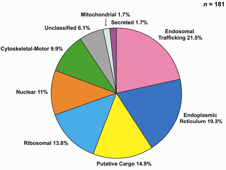
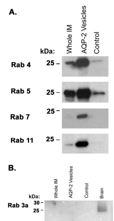
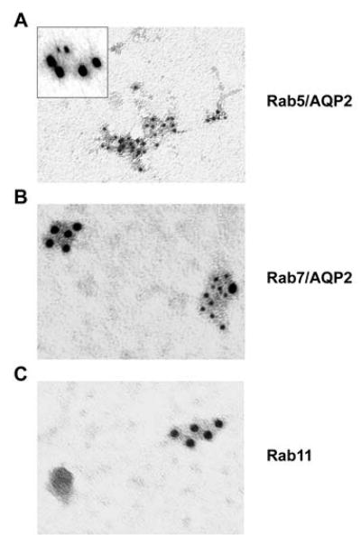
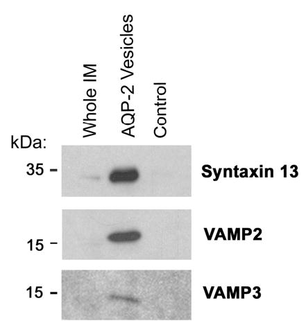
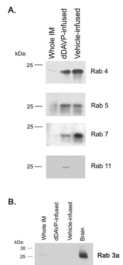
Vasopressin acts on renal collecting duct cells to stimulate translocation of aquaporin-2 (AQP2)-containing membrane vesicles from throughout the cytoplasm to the apical region. The vesicles fuse with the plasma membrane to increase water permeability. To identify the intracellular membrane compartments that contain AQP2, we carried out LC-MS/MS-based proteomic analysis of immunoisolated AQP2-containing intracellular vesicles from rat inner medullary collecting duct. Immunogold electron microscopy and immunoblotting confirmed heavy AQP2 labeling of immunoisolated vesicles. Vesicle proteins were separated by SDS-PAGE followed by in-gel trypsin digestion in consecutive gel slices and identification by LC-MS/MS. Identification of Rab GTPases 4, 5, 18, and 21 (associated with early endosomes); Rab7 (late endosomes); and Rab11 and Rab25 (recycling endosomes) indicate that a substantial fraction of intracellular AQP2 is present in endosomal compartments. In addition, several endosome-associated SNARE proteins were identified including syntaxin-7, syntaxin-12, syntaxin-13, Vti1a, vesicle-associated membrane protein 2, and vesicle-associated membrane protein 3. Rab3 was not found, however, either by mass spectrometry or immunoblotting, suggesting a relative lack of AQP2 in secretory vesicles. Additionally, we identified markers of the trans-Golgi network, components of the exocyst complex, and several motor proteins including myosin 1C, non-muscle myosins IIA and IIB, myosin VI, and myosin IXB. Beyond this, identification of multiple endoplasmic reticulum-resident proteins and ribosomal proteins indicated that a substantial fraction of intracellular AQP2 is present in rough endoplasmic reticulum. These results show that AQP2-containing vesicles are heterogeneous and that intracellular AQP2 resides chiefly in endosomes, trans-Golgi network, and rough endoplasmic reticulum.


Figures

Figure 1
Figure 1
Flow diagrams for (A) immunoisolation of intracellular AQP2 vesicles and (B) proteomic analysis of AQP2 vesicles.
Figure 2
Figure 2. A) Immunoblot characterization of biotinylated chicken anti-AQP2 antibody
The biotinylated chicken anti-AQP2 (right panel) recognized nonglycosylated AQP2 at 29 kDa and glycosylated AQP2 at 35–40 kDa, as seen with previously characterized (13) rabbit anti-AQP2 antibody (left panel). Immunoblot using biotinylated chicken antibody utilized streptavidin-horseradish peroxidase (HRP) conjugate in lieu of HRP-conjugated secondary antibody. B) Surface biotinylation showing relative absence of plasma membrane proteins in low-density membrane fraction. Surface biotinylation of inner medullary collecting duct (IMCD) suspension was performed (see Methods). The IMCD cells were homogenized and then subjected to differential centrifugation to obtain 17,000 Xg pellet (high-density membrane fraction) and 200,000 Xg pellet (low-density membrane fraction). Detection of biotinylated proteins in these two fractions was accomplished by SDS-PAGE followed by electroblotting and probing the blots with a streptavidin-horseradish peroxidase (HRP) conjugate. Signal was detected through use of chemiluminescence and light-sensitive film. Three μg of protein was loaded in both lanes. The two lanes shown are from the same exposure of the same blot.
Figure 3
Figure 3
(A) Coomassie-stained gels of immunoisolated proteins and proteins isolated nonspecifically in non-immune IgY control. 1-D SDS-PAGE using 12% polyacrylamide. Heavy band in control lane is BSA. Gel was sliced as indicated to obtain 35 gel pieces for in-gel trypsinization and LC-MS/MS analysis. (B) Immunoblot of AQP2 in the whole renal inner medulla homogenate, AQP2-immunoisolated vesicles, and control sample. Each lane was loaded with an equal amount of total protein. The immunoblot was probed with a rabbit anti-AQP2 antibody. Film was deliberately overexposed to visualize weak AQP2 signal in control lane. AQP2 was markedly enriched in AQP2-immunoisolated vesicles as compared to the control sample. (C) Immuno-gold electron microscopy of immunoisolated vesicles. Primary antibody was affinity purified rabbit anti-AQP2.
Figure 4
Figure 4. Classification of proteins identified with two or more unique peptides in immunoisolated AQP2-vesicles by LC-MS/MS analysis
Protein classification utilized Rat Genome Database (http://rgd.mcw.edu/) and Harvester (EMBL, http://harvester.embl.de/).
Figure 5
Figure 5. Confirmatory immunoblots for Rab GTPases
(A) Immunoblots for endosome-associated Rab GTPases in AQP2-immunoisolated vesicles: Rab4 (present in early endosomes), Rab5 (present in early endosomes and clathrin-coated endocytic vesicles), Rab7 (present in late endosomes), and Rab11 (present in recycling endosomes). (B) Immunoblot comparing abundance of Rab3a in the whole renal inner medulla homogenate, AQP2-immunoisolated vesicles, non-immune IgY control sample, and whole brain homogenate.
Figure 6
Figure 6. Electron micrographs showing immuno-gold labeling of AQP2-immunoisolated vesicles
(A) Rab5 (inset, Rab5 [small particles] and AQP2 [large particles], (B) Rab7 (small gold particles) and AQP2 (large gold particles), (c) Rab11 (single labeling only).
Figure 7
Figure 7
Immunoblot using anti-ARF 6 confirming presence of ARF 6 in AQP2-immunoisolated vesicles.
Figure 8
Figure 8
Immunoblots confirming presence of SNARE proteins syntaxin 13, VAMP2 (synaptobrevin II), and VAMP3 (cellubrevin) in AQP2-immunoisolated vesicles.
Figure 9
Figure 9
Immunoblots confirming presence of myosin 1C, myosin IIA, and myosin VI in AQP2-immunoisolated vesicles.
Figure 10
Figure 10
Immunoblot using anti-ubiquitin antibody. Note smear over a molecular weight range from 25 kDa to over 250 kDa, suggesting the presence of a variety of ubiquitinated proteins in AQP2-immunoisolated vesicles.
Figure 11
Figure 11
Immunoblots identifying exocyst complex proteins Sec 8, Sec 6, and Ral A in AQP2-immunoisolated vesicles.
Figure 12
Figure 12. Immunoblots showing relative abundance of various Rab GTPases in AQP2-immunoisolated intracellular vesicles from Brattleboro rats treated with either dDAVP or vehicle
(A) Endosome-associated Rab GTPases, including Rab4 and 5 (early endosomes), Rab7 (late endosomes), and Rab11 (recycling endosomes). (B) Rab3a (normally associated with secretory vesicles).

References

    1. Nielsen S, Chou CL, Marples D, Christensen EI, Kishore BK, Knepper MA. Proceedings of the National Academy of Sciences, USA. 1995;92:1013–1017. - PMC - PubMed
    1. Nielsen S, DiGiovanni SR, Christensen EI, Knepper MA, Harris HW. Proceedings of the National Academy of Sciences, USA. 1993;90:11663–11667. - PMC - PubMed
    1. van Balkom BW, Graat MP, van Raak M, Hofman E, van der SP, Deen PM. AmJPhysiol Cell Physiol. 2004;286:C372–C379. - PubMed
    1. Lu H, Sun TX, Bouley R, Blackburn K, McLaughlin M, Brown D. AmJPhysiol Renal Physiol. 2004;286:F233–F243. - PubMed
    1. Asai T, Kuwahara M, Kurihara H, Sakai T, Terada Y, Marumo F, Sasaki S. Kidney Int. 2003;64:2–10. - PubMed

Publication types

LinkOut - more resources