

Structure and function of parallel pathways in the primate early visual system
- PMID: 15905213
- PMCID: PMC1464718
- DOI: 10.1113/jphysiol.2005.088047
Structure and function of parallel pathways in the primate early visual system
Abstract
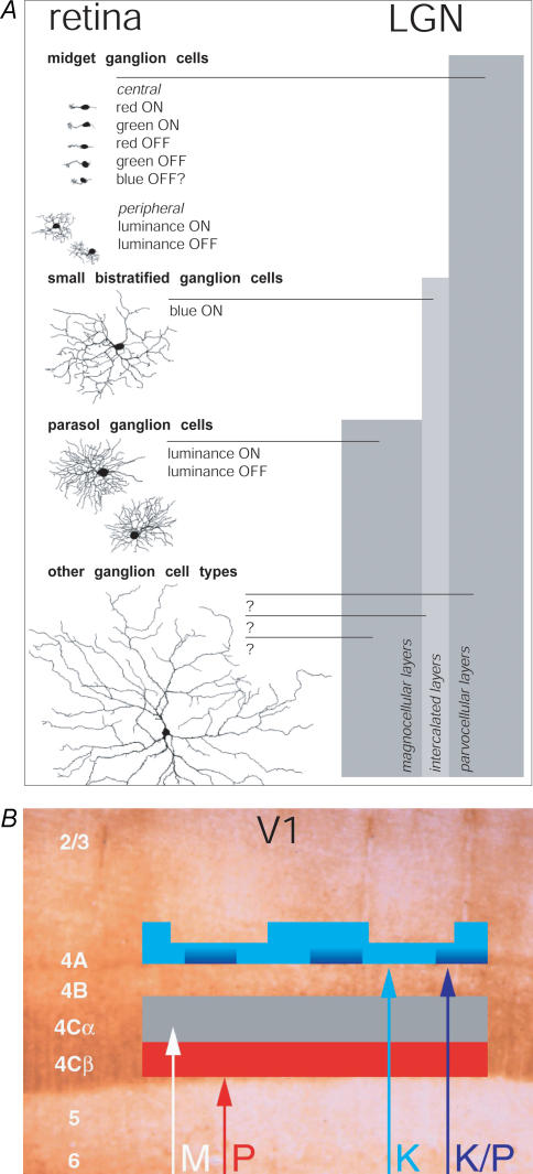
Parallel processing streams in the primate visual system originate from more than a dozen anatomically and functionally distinct types of retinal ganglion cells (RGCs). A central problem in determining how visual information is processed is understanding how each of these RGC types connects to more central structures, including the lateral geniculate nucleus (LGN) of the thalamus and (via the LGN) the primary visual cortex. Nevertheless, the available functional and anatomical evidence linking together specific cell types across these structures is surprisingly indirect. This review evaluates the available evidence and assesses the strength of the many inferences that can be made from these observations. There is strong evidence that parasol RGCs are the provenance of the magnocellular (M) visual pathway and that midget RGCs give rise to the parvocellular (P) pathway. Furthermore, the M and P pathways remain segregated up to the input layer of primary visual cortex. The relationships between the numerous other RGC types and cell types in the LGN remain less certain. and there remains ambiguity about how best to define additional pathways, such as the koniocellular (K) pathway, which probably arise from these other, less common, RGC types.
Figures
Comment in
-
The senses.J Physiol. 2005 Jul 1;566(Pt 1):5. doi: 10.1113/jphysiol.2005.090837. Epub 2005 May 26. J Physiol. 2005. PMID: 15919707 Free PMC article. No abstract available.
References
-
- Ahmad KM, Klug K, Herr S, Sterling P, Schein S. Cell density ratios in a foveal patch in macaque retina. Vis Neurosci. 2003;20:189–209. - PubMed
-
- Beaulieu C, Kisvarday Z, Somogyi P, Cynader M, Cowey A. Quantitative distribution of GABA-immunopositive and -immunonegative neurons and synapses in the monkey striate cortex (area 17) Cereb Cortex. 1992;2:295–309. - PubMed
-
- Casagrande VA. A third parallel visual pathway to primate area V1. Trends Neurosci. 1994;17:305–310. - PubMed
Publication types
MeSH terms
LinkOut - more resources
Full Text Sources