Review
. 2005 Aug 1;566(Pt 3):645-56.
doi: 10.1113/jphysiol.2005.086405. Epub 2005 May 19.

Rhythmicity in arterial smooth muscle

Affiliations
Review

Rhythmicity in arterial smooth muscle

Rebecca E Haddock et al. J Physiol. .

Abstract

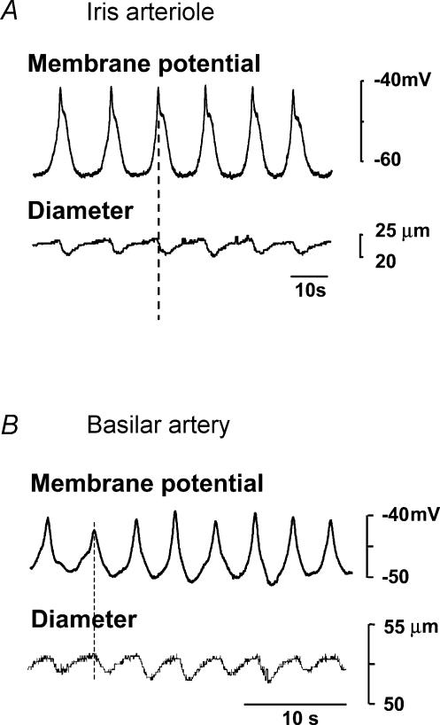
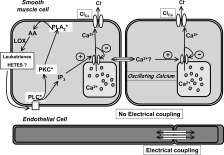
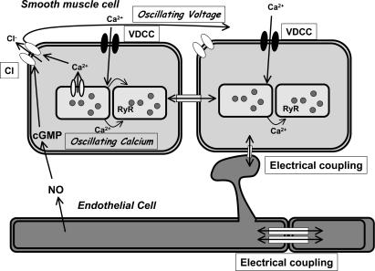
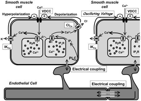
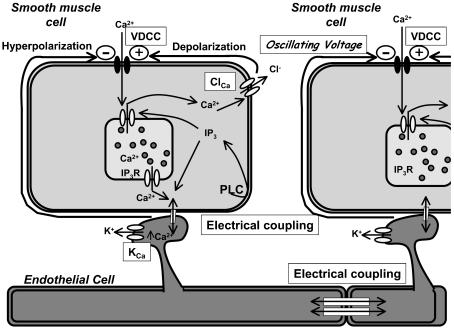
Many arteries and arterioles exhibit rhythmical contractions which are synchronous over considerable distances. This vasomotion is likely to assist in tissue perfusion especially during periods of altered metabolism or perfusion pressure. While the mechanism underlying vascular rhythmicity has been investigated for many years, it has only been recently, with the advent of imaging techniques for visualizing intracellular calcium release, that significant advances have been made. These methods, when combined with mechanical and electrophysiological recordings, have demonstrated that the rhythm depends critically on calcium released from intracellular stores within the smooth muscle cells and on cell coupling via gap junctions to synchronize oscillations in calcium release amongst adjacent cells. While these factors are common to all vessels studied to date, the contribution of voltage-dependent channels and the endothelium varies amongst different vessels. The basic mechanism for rhythmical activity in arteries thus differs from its counterpart in non-vascular smooth muscle, where specific networks of pacemaker cells generate electrical potentials which drive activity within the otherwise quiescent muscle cells.


Figures

Figure 1
Figure 1. Membrane potential oscillations underlying vasomotion in rat irideal arterioles and basilar arteries
While depolarizations precede constrictions in both vessels, the most negative membrane potential reached in the iris is less than −60 mV while that in the basilar is considerably more positive and less stable. Note also that the oscillations in the iris are slower than those in the basilar artery. Part B reproduced with permission of Blackwell Publishing from Haddock & Hill (2002).
Figure 2
Figure 2. The voltage-independent coupled oscillator in iris arterioles
Constitutive activity of phospholipase C (PLC) leads to the production of IP3 and basal release of Ca2+ from the IP3-sensitive Ca2+ store. Simultaneous activation of protein kinase C (PKC) stimulates phospholipase A2 (PLA2) and breakdown of arachidonic acid via the lipoxygenase (LOX) pathway. The resultant metabolites further stimulate the PLC pathway resulting in cyclical oscillations in [Ca2+]i due to the biphasic regulation of the IP3 receptor by Ca2+. This also produces coincident cyclical depolarizations of the cell membrane through opening of a Ca2+-dependent chloride channel (ClCa). Synchronization results from passage of Ca2+ through gap junctions. Voltage-dependent Ca2+ channels are not activated.
Figure 3
Figure 3. The voltage-dependent coupled oscillator in rat small mesenteric arteries
Application of agonist induces Ca2+ release from intracellular stores. This is oscillatory and, in combination with cGMP, produced by the action of endothelially derived nitric oxide (NO), activates a Ca2+- and cGMP-dependent chloride channel (Cl) which intermittently depolarizes the membrane potential. If sufficient cells depolarize together the depolarization opens voltage-dependent Ca2+ channels (VDCC) causing extracellular Ca2+ influx into the cells. As the cells are all electrically coupled, this Ca2+ entry is simultaneous and it synchronizes the intermittent Ca2+ release from ryanodine receptors (RyR) in adjacent cells.
Figure 4
Figure 4. The voltage-dependent membrane oscillator in rat basilar arteries
Constitutive activity of phospholipase C (PLC) leads to the production of IP3 and release of Ca2+ to activate a Ca2+-dependent chloride channel (ClCa). This current depolarizes the membrane potential and opens voltage-dependent Ca2+ channels (VDCC) causing extracellular Ca2+ influx. This Ca2+ influx activates ryanodine receptors (RyRs) causing further Ca2+ release which opens intermediate conductance KCa channels and the membrane hyperpolarizes, thereby closing the VDCCs. Coupling between adjacent smooth muscle cells is poor but the oscillating membrane voltage acts to synchronize adjacent cells due to electrical coupling via the endothelium. Closure of the VDCCs following SMC hyperpolarization ensures the vasodilatory part of the cycle while the tonic depolarization through the ClCa channels ensures that the VDCCs reopen and the cycle oscillates.
Figure 5
Figure 5. The voltage-dependent membrane oscillator in rat mesenteric arteries
Application of agonist induces Ca2+ release from intracellular stores and activation of a depolarizing current. The resulting Ca2+ influx through VDCCs synchronizes and augments release from intracellular stores. Either Ca2+ or IP3 passes through myoendothelial gap junctions into the endothelial cells to activate KCa channels. The ensuing hyperpolarization is then transferred electrotonically back into the SMCs where it reduces Ca2+ entry due to the closure of VDCCs.

References

    1. Bartlett IS, Crane GJ, Neild TO, Segal SS. Electrophysiological basis of arteriolar vasomotion in vivo. J Vasc Res. 2000;37:568–575. - PubMed
    1. Beech DJ. Emerging functions of 10 types of transient receptor potential (TRP) cationic channel in vascular smooth muscle. Proc Aust Physiol Soc. 2005;36:1–8. - PMC - PubMed
    1. Benham CD, Bolton TB. Spontaneous transient outward currents in single visceral and vascular smooth muscle cells of the rabbit. J Physiol. 1986;381:385–406. - PMC - PubMed
    1. Bolton TB, Gordienko DV, Povstyan OV, Harhun MI, Pucovsky V. Smooth muscle cells and interstitial cells of blood vessels. Cell Calcium. 2004;35:643–657. - PubMed
    1. Bonnet S, Belus A, Hyvelin JM, Roux E, Marthan R, Savineau JP. Effect of chronic hypoxia on agonist-induced tone and calcium signaling in rat pulmonary artery. Am J Physiol Lung Cell Mol Physiol. 2001;281:L193–L201. - PubMed

LinkOut - more resources