. 2005 Jun 1;174(11):7285-91.
doi: 10.4049/jimmunol.174.11.7285.

Mast cell protease 5 mediates ischemia-reperfusion injury of mouse skeletal muscle

Affiliations

Mast cell protease 5 mediates ischemia-reperfusion injury of mouse skeletal muscle

J Pablo Abonia et al. J Immunol. .

Abstract

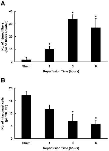
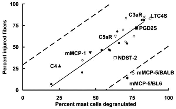
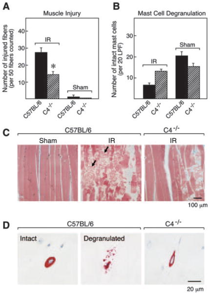
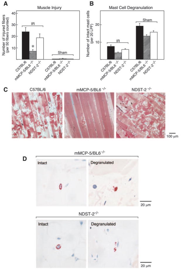
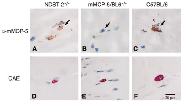
Ischemia with subsequent reperfusion (IR) injury is a significant clinical problem that occurs after physical and surgical trauma, myocardial infarction, and organ transplantation. IR injury of mouse skeletal muscle depends on the presence of both natural IgM and an intact C pathway. Disruption of the skeletal muscle architecture and permeability also requires mast cell (MC) participation, as revealed by the fact that IR injury is markedly reduced in c-kit defective, MC-deficient mouse strains. In this study, we sought to identify the pathobiologic MC products expressed in IR injury using transgenic mouse strains with normal MC development, except for the lack of a particular MC-derived mediator. Histologic analysis of skeletal muscle from BALB/c and C57BL/6 mice revealed a strong positive correlation (R(2) = 0.85) between the extent of IR injury and the level of MC degranulation. Linkage between C activation and MC degranulation was demonstrated in mice lacking C4, in which only limited MC degranulation and muscle injury were apparent. No reduction in injury was observed in transgenic mice lacking leukotriene C(4) synthase, hemopoietic PGD(2) synthase, N-deacetylase/N-sulfotransferase-2 (enzyme involved in heparin biosynthesis), or mouse MC protease (mMCP) 1. In contrast, muscle injury was significantly attenuated in mMCP-5-null mice. The MCs that reside in skeletal muscle contain abundant amounts of mMCP-5 which is the serine protease that is most similar in sequence to human MC chymase. We now report a cytotoxic activity associated with a MC-specific protease and demonstrate that mMCP-5 is critical for irreversible IR injury of skeletal muscle.


Conflict of interest statement

Disclosures: The authors have no financial conflict of interest.

Figures

FIGURE 1
FIGURE 1
Assessment of the time-dependent appearance of muscle injury and MC degranulation. Histological assessment of muscle injury (A) and MC degranulation (B) after 2 h of ischemia and various times of reperfusion in C57BL/6 mice. A, The mean numbers (±SEM) of injured fibers per 50 fc from 3 to 6 animals in three separate experiments are plotted (*, p < 0.05 relative to sham). B, The mean numbers of intact MC (±SEM) in 20 LPF are plotted for the same animals described in A (*, p < 0.05 relative to sham).
FIGURE 2
FIGURE 2
Correlation of IR-induced muscle damage with MC degranulation. The mean percentage of injured muscle fibers vs the mean percentage of degranulated MCs after hind limb IR injury was plotted for each mouse strain studied in this investigation. The correlation coefficient and solid trend line are based on the mean values from the injured wild-type control groups (C57BL/6 (●), n = 10, and BALB/c (○), n = 5) in the individual experiments including the time course presented in Fig. 1. The Pearson correlation coefficient (R2) for the data for wild-type mice alone is 0.85. The 95% confidence interval relative to the trend line is indicated by dashed lines. Also indicated are the mean percentages for muscle injury and MC degranulation obtained from all studies with transgenic mice that do not express C3aR (*), C5aR (∇), C4 (▲), LTC4S (△), PGD2S (■), mMCP-1 (▼), mMCP-5/BL6 (◆), mMCP-5/BALB (◇), or NDST-2 (□).
FIGURE 3
FIGURE 3
IR-induced muscle damage and MC degranulation in C4−/− mice. A, The mean number (±SEM) of injured muscle fibers per 50 fc is shown for 8 IR-treated C57BL/6 mice, 12 IR-treated C4−/− mice, 3 sham-treated C57BL/6 mice, and 4 sham-treated C4−/− mice in two separate experiments (*, p < 0.01 relative to wild type). B, The mean number (±SEM) of intact MCs per 20 LPF was quantitated for each group described in A. C and D, Representative histological sections from these experiments. C, Gastrocnemius muscle sections were stained with Masson's trichrome. Large numbers of injured muscle fibers were detected in the muscle of IR-treated C57BL/6 mice with serpigenous strands of actin/myosin and rhabdomyolysis (arrows, middle panel). Minimal irreversible damage occurred in sham-treated C57BL/6 mice (left panel) and IR-treated C4−/− mice (right panel). D, High-power (×50 objective) magnification fields of the chloroacetate esterase-positive MCs in the muscle of the three groups as in C. Many exocytosed granules were found in the extracellular milieu adjacent to the activated MCs in IR-treated C57BL/6 mice (middle panel). These exocytosed granules were rarely seen in sham-treated C57BL/6 mice (left panel) or IR-treated C4−/− mice (right panel).
FIGURE 4
FIGURE 4
IR-induced muscle damage and MC degranulation in mMCP-5/BL6−/− and in NDST-2−/− mice. A, The mean number (±SEM) of injured muscle fibers per 50 fc is shown for 11 IR-treated C57BL/6 mice, 12 IR-treated mMCP-5/BL6−/− mice, 6 IR-treated NDST-2−/− mice, 8 sham-treated C57BL/6 mice, 8 sham-treated mMCP-5/BL6−/− mice, and 3 sham-treated NDST-2−/− mice (*, p < 0.01). B, The mean number (±SEM) of intact MCs per 20 LPF were quantitated for each group. C, Representative histological sections from IR-treated C57BL/6 (left panel), mMCP-5/BL6−/− (middle panel), and NDST-2−/− (right panel) mice are shown. Extensive muscle damage (including rhabdomyolysis) occurs in IR-treated C57BL/6 mice and NDST-2−/− mice but not in mMCP-5/BL6−/− mice. D, High-power magnification fields of the chloroacetate esterase-positive MCs in the muscle of the mMCP-5/BL6−/− and NDST-2−/− mice. Many exocytosed granules were found in the extracellular milieu adjacent to the activated MCs in IR-treated mMCP-5/BL6−/− mice and NDST-2−/− mice but not in their sham-treated controls.
FIGURE 5
FIGURE 5
Immunohistochemical detection of mMCP-5 in NDST-2−/− and in C57BL/6 mice but not in mMCP-5/BL6−/− mice. AC, The presence or absence of mMCP-5 in MCs was evaluated in frozen sections of skeletal muscle from NDST-2−/− (A), mMCP-5/BL6−/− (B), or wild-type C57BL/6 (C) mice using anti-mMCP-5. MCs are indicted by arrowheads. DF, The size of the skeletal muscle MCs in the various mouse strains was compared using chloroacetate esterase (CAE) reactivity to identify the cells in paraformaldehyde-fixed sections of skeletal muscle from NDST-2−/− (D), mMCP-5/BL6−/− (E), or wild-type C57BL/6 (F) mice.

References

    1. Weiser MR, Williams JP, Moore FD, Jr, Kobzik L, Ma M, Hechtman HB, Carroll MC. Reperfusion injury of ischemic skeletal muscle is mediated by natural antibody and complement. J Exp Med. 1996;183:2343–2348. - PMC - PubMed
    1. Kyriakides C, Austen W, Jr, Wang Y, Favuzza J, Kobzik L, Moore FD, Jr, Hechtman HB. Skeletal muscle reperfusion injury is mediated by neutrophils and the complement membrane attack complex. Am J Physiol. 1999;277:C1263–C1268. - PubMed
    1. Lazarus B, Messina A, Barker JE, Hurley JV, Romeo R, Morrison WA, Knight KR. The role of mast cells in ischaemia-reperfusion injury in murine skeletal muscle. J Pathol. 2000;191:443–448. - PubMed
    1. Mukundan C, Gurish MF, Austen KF, Hechtman HB, Friend DS. Mast cell mediation of muscle and pulmonary injury following hindlimb ischemia-reperfusion. J Histochem Cytochem. 2001;49:1055–1056. - PubMed
    1. Bortolotto SK, Morrison WA, Han X, Messina A. Mast cells play a pivotal role in ischaemia reperfusion injury to skeletal muscles. Lab Invest. 2004;84:1103–1111. - PubMed

Publication types

MeSH terms