

doi: 10.1002/prot.20410.
1.6 A crystal structure of YteR protein from Bacillus subtilis, a predicted lyase
Affiliations
- PMID: 15906318
- PMCID: PMC2792013
- DOI: 10.1002/prot.20410
Item in Clipboard
1.6 A crystal structure of YteR protein from Bacillus subtilis, a predicted lyase
Proteins.
.
No abstract available
Figures
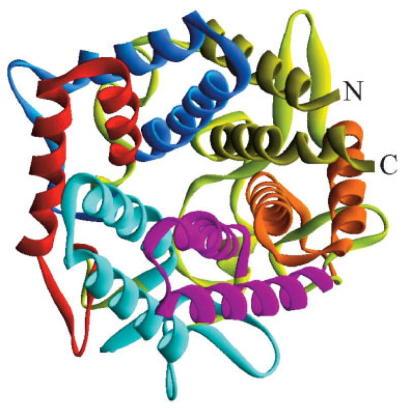
This ribbon drawing shows the closed circular array formed by the six-alpha hairpins of YteR, each indicated by a unique color. Counterclockwise from the N-terminus: HP1 is blue, HP2 is red, HP3 is light blue, HP4 is purple, HP5 is orange, and H6 is yellow-green. The picture was generated using DS ViewerPro.
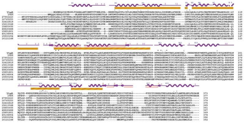
Multiple sequence alignment, superfamily motifs, and secondary structure analysis of YteR: the alignment of one-letter amino acid sequences of Yter and 11 homologous structures was performed by ClusterX. The numbers in the left column are NCBI gi accession codes, and the numbers in the right column indicate the residue numbers at the end of each row. The yellow bars designate residues in YteR that match the six-hairpin glycosyltransferases motif 48208 (Typ41 to Asp88, Ala92 to Lys133, Trp141 to Cys206, Arg270 to Lys289, Asp297 to Lys315). The secondary elements are indicated above the one-letter amino acid codes of YteR.
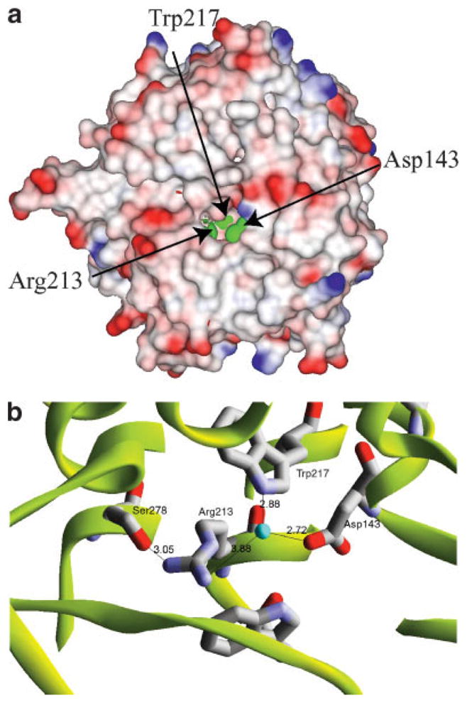
(a) Electrostatic surface potentials of YteR protein. The green represents solvent-exposed putative active site residues. The surface was calculated with 1.4 Å radius. Blue shading represents positive potential and red the negative potential. Labels indicate amino acid residues. This image was created by the program DS ViewerPro. (b) Close-up view of the putative active site. Arg213, Trp217, Asp143, and Ser278 are coordinated by ordered water molecule. These residues are located close enough for hydrogen bonds. Atomic distances in angstroms are labeled in black. This image was created by the program DS ViewerPro.
References
-
- Holm L, Sander C. Protein structure comparison by alignment of distance matrices. J Mol Biol. 1993;233:123–138. - PubMed
-
- Murzin AG, Brenner SE, Hubbard T, Chothia C. SCOP: a structural classification of proteins database for the investigation of sequences and structures. J Mol Biol. 1995;247:536–540. - PubMed
-
- Walsh MA, Dementieva I, Evans G, Sanishvili R, Joachimiak A. Taking MAD to the extreme: ultrafast protein structure determination. Acta Crystallogr D Biol Crystallogr. 1999;55:1168–1173. - PubMed
Publication types
MeSH terms
Substances
Associated data
- Actions
- Actions
- Actions
- Actions
- Actions
- Actions
- Actions
Grants and funding
LinkOut - more resources
Full Text Sources
Molecular Biology Databases
