

Solitary intra-abdominal tuberculous lymphadenopathy mimicking duodenal GIST
- PMID: 15906957
- PMCID: PMC3891416
- DOI: 10.3904/kjim.2005.20.1.72
Solitary intra-abdominal tuberculous lymphadenopathy mimicking duodenal GIST
Abstract
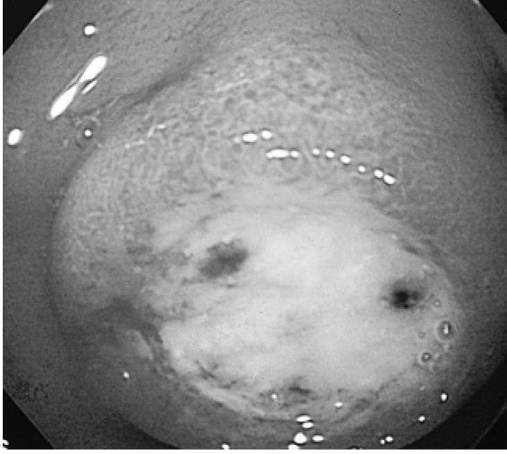
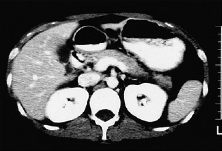
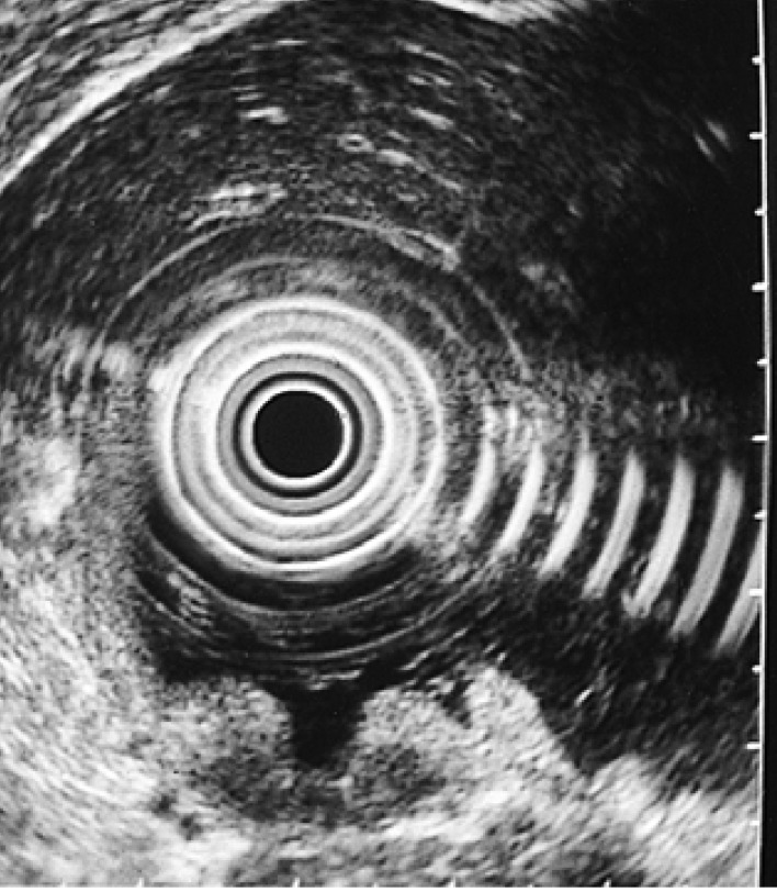
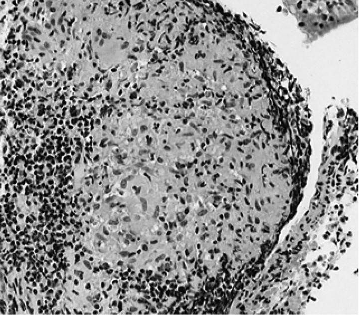
Tuberculosis remains prevalent in developing countries and has recently re-emerged in the Western world. Intra-abdominal tuberculosis can mimic a variety of other abdominal disorders, and here we describe a patient with solitary tuberculous mesenteric lymphadenopathy mimicking duodenal gastrointestinal stromal tumor (GIST). A 22-year-old woman complained of epigastric discomfort and was presumed to have a duodenal GIST after an endoscopic examination and abdominal CT scan. However, exploratory laparotomy revealed an enlarged node penetrating the duodenal bulb, which was diagnosed histopathologically as tuberculous lymphadenitis. This case suggests that in regions with a high prevalence of tuberculosis, intra-abdominal tuberculosis is often mistaken as a malignant neoplasm. A high index of suspicion and the accurate nonsurgical diagnosis of intra-abdominal tuberculosis continues to be a challenge.
Figures
Similar articles
-
Peripancreatic tuberculous lymphadenitis mimicking carcinoma: report of a case.Acta Chir Belg. 2004 Jun;104(3):338-40. doi: 10.1080/00015458.2004.11679568. Acta Chir Belg. 2004. PMID: 15285551
-
Tuberculous mesenteric lymphadenitis mimicking pancreatic carcinoma.Hepatogastroenterology. 1996 Nov-Dec;43(12):1653-5. Hepatogastroenterology. 1996. PMID: 8975983
-
[Upper gastrointestinal bleeding in an 81-year-old woman].Gastroenterol Hepatol. 2010 Jan;33(1):30-2. doi: 10.1016/j.gastrohep.2009.07.011. Epub 2009 Oct 9. Gastroenterol Hepatol. 2010. PMID: 19819044 Spanish.
-
Diagnosis of abdominal tuberculosis: experience from 11 cases and review of the literature.World J Gastroenterol. 2004 Dec 15;10(24):3647-9. doi: 10.3748/wjg.v10.i24.3647. World J Gastroenterol. 2004. PMID: 15534923 Free PMC article. Review.
-
[A case of tuberculous mesenteric lymphadenitis detected by abdominal symptom after 4 months' antituberculous chemotherapy against pulmonary tuberculosis].Kekkaku. 1991 Aug;66(8):543-51. Kekkaku. 1991. PMID: 1921095 Review. Japanese.
Cited by
-
Splenic artery aneurysm presenting with clinical features of a bleeding gastric gastrointestinal stromal tumour.J Surg Case Rep. 2011 Jun 1;2011(6):1. doi: 10.1093/jscr/2011.6.1. J Surg Case Rep. 2011. PMID: 24949696 Free PMC article.
-
Abdominal cyst of unclear aetiology: gastrointestinal stromal tumour or reactivation of abdominal tuberculosis.BMJ Case Rep. 2022 Jan 6;15(1):e245767. doi: 10.1136/bcr-2021-245767. BMJ Case Rep. 2022. PMID: 34992056 Free PMC article.
-
The spectrum of abdominal tuberculosis in a developed country: a single institution's experience over 7 years.J Gastrointest Surg. 2009 Jan;13(1):142-7. doi: 10.1007/s11605-008-0669-6. Epub 2008 Sep 3. J Gastrointest Surg. 2009. PMID: 18769984
-
Left-sided portal hypertension caused by peripancreatic lymph node tuberculosis misdiagnosed as pancreatic cancer: a case report and literature review.BMC Gastroenterol. 2020 Aug 18;20(1):276. doi: 10.1186/s12876-020-01420-x. BMC Gastroenterol. 2020. PMID: 32811429 Free PMC article. Review.
-
Neoplasm-like abdominal nonhematogenous disseminated tuberculous lymphadenopathy: CT evaluation of 12 cases and literature review.World J Gastroenterol. 2011 Sep 21;17(35):4038-43. doi: 10.3748/wjg.v17.i35.4038. World J Gastroenterol. 2011. PMID: 22046094 Free PMC article.
References
-
- Rieder HL, Cauthen GM, Kelly GD, Bloch AB, Snider DE., Jr Tuberculosis in the United States. JAMA. 1989;262:385–389. - PubMed
-
- Bargallo N, Nicolau C, Luburich P, Ayuso C, Cardenal C, Gimeno F. Intestinal tuberculosis in AIDS. Gastrointest Radiol. 1992;17:115–118. - PubMed
-
- Marshall JB. Tuberculosis of the gastrointestinal tract and peritoneum. Am J Gastroenterol. 1993;88:989–999. - PubMed
-
- Jakubowski A, Elwood RK, Enarson DA. Clinical features of abdominal tuberculosis. J Infect Dis. 1988;158:687–692. - PubMed
-
- al Karawi MA, Mohamed AE, Yasawy MI, Graham DY, Shariq S, Ahmed AM, al Jumah A, Ghandour Z. Protean manifestation of gastrointestinal tuberculosis: report on 130 patients. J Clin Gastroenterol. 1995;20:225–232. - PubMed
Publication types
MeSH terms
LinkOut - more resources
Full Text Sources
Medical