Identification, characterization and metagenome analysis of oocyte-specific genes organized in clusters in the mouse genome
- PMID: 15907208
- PMCID: PMC1166550
- DOI: 10.1186/1471-2164-6-76
Identification, characterization and metagenome analysis of oocyte-specific genes organized in clusters in the mouse genome
Abstract
Background: Genes specifically expressed in the oocyte play key roles in oogenesis, ovarian folliculogenesis, fertilization and/or early embryonic development. In an attempt to identify novel oocyte-specific genes in the mouse, we have used an in silico subtraction methodology, and we have focused our attention on genes that are organized in genomic clusters.
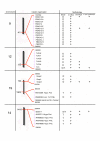
Results: In the present work, five clusters have been studied: a cluster of thirteen genes characterized by an F-box domain localized on chromosome 9, a cluster of six genes related to T-cell leukaemia/lymphoma protein 1 (Tcl1) on chromosome 12, a cluster composed of a SPErm-associated glutamate (E)-Rich (Speer) protein expressed in the oocyte in the vicinity of four unknown genes specifically expressed in the testis on chromosome 14, a cluster composed of the oocyte secreted protein-1 (Oosp-1) gene and two Oosp-related genes on chromosome 19, all three being characterized by a partial N-terminal zona pellucida-like domain, and another small cluster of two genes on chromosome 19 as well, composed of a TWIK-Related spinal cord K+ channel encoding-gene, and an unknown gene predicted in silico to be testis-specific. The specificity of expression was confirmed by RT-PCR and in situ hybridization for eight and five of them, respectively. Finally, we showed by comparing all of the isolated and clustered oocyte-specific genes identified so far in the mouse genome, that the oocyte-specific clusters are significantly closer to telomeres than isolated oocyte-specific genes are.
Conclusion: We have studied five clusters of genes specifically expressed in female, some of them being also expressed in male germ-cells. Moreover, contrarily to non-clustered oocyte-specific genes, those that are organized in clusters tend to map near chromosome ends, suggesting that this specific near-telomere position of oocyte-clusters in rodents could constitute an evolutionary advantage. Understanding the biological benefits of such an organization as well as the mechanisms leading to a specific oocyte expression in these clusters now requires further investigation.
Figures


References
-
- Varani S, Matzuk MM. Phenotypic effects of knockout of oocyte-specific genes. Ernst Schering Res Found Workshop. 2002:63–79. - PubMed
-
- Galloway SM, McNatty KP, Cambridge LM, Laitinen MP, Juengel JL, Jokiranta TS, McLaren RJ, Luiro K, Dodds KG, Montgomery GW, Beattie AE, Davis GH, Ritvos O. Mutations in an oocyte-derived growth factor gene (BMP15) cause increased ovulation rate and infertility in a dosage-sensitive manner. Nat Genet. 2000;25:279–283. doi: 10.1038/77033. - DOI - PubMed
MeSH terms
Substances
LinkOut - more resources
Full Text Sources
Molecular Biology Databases
Research Materials

