. 2005 May 20;121(4):607-620.
doi: 10.1016/j.cell.2005.03.012.

The v-ATPase V0 subunit a1 is required for a late step in synaptic vesicle exocytosis in Drosophila

Affiliations

The v-ATPase V0 subunit a1 is required for a late step in synaptic vesicle exocytosis in Drosophila

P Robin Hiesinger et al. Cell. .

Abstract

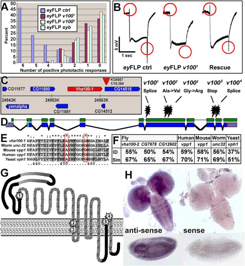
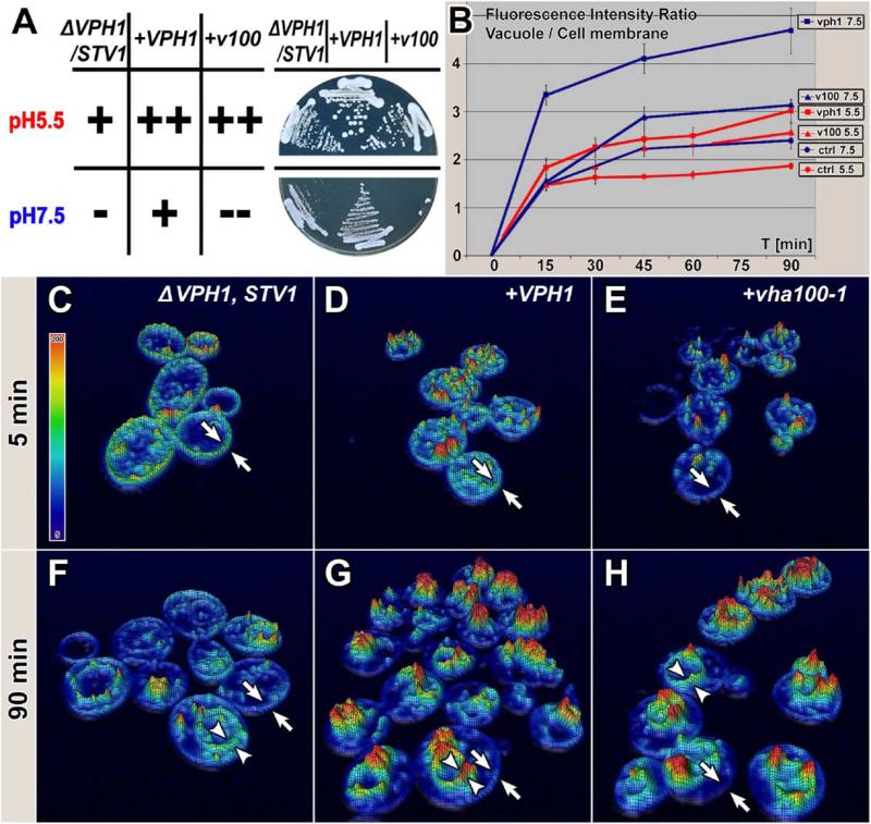
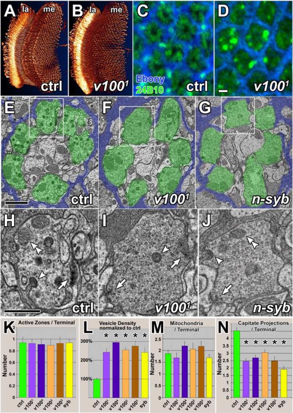
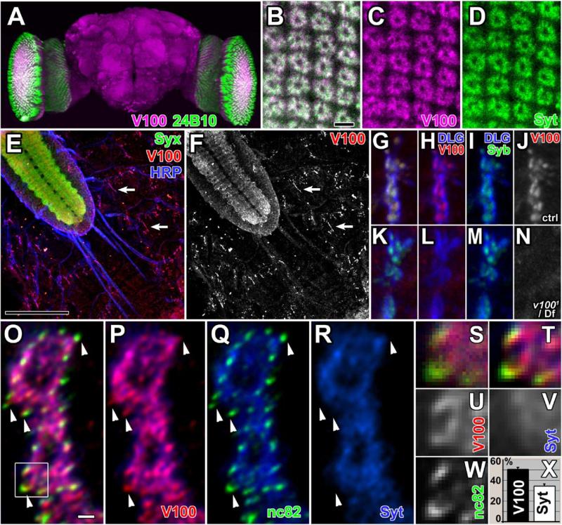
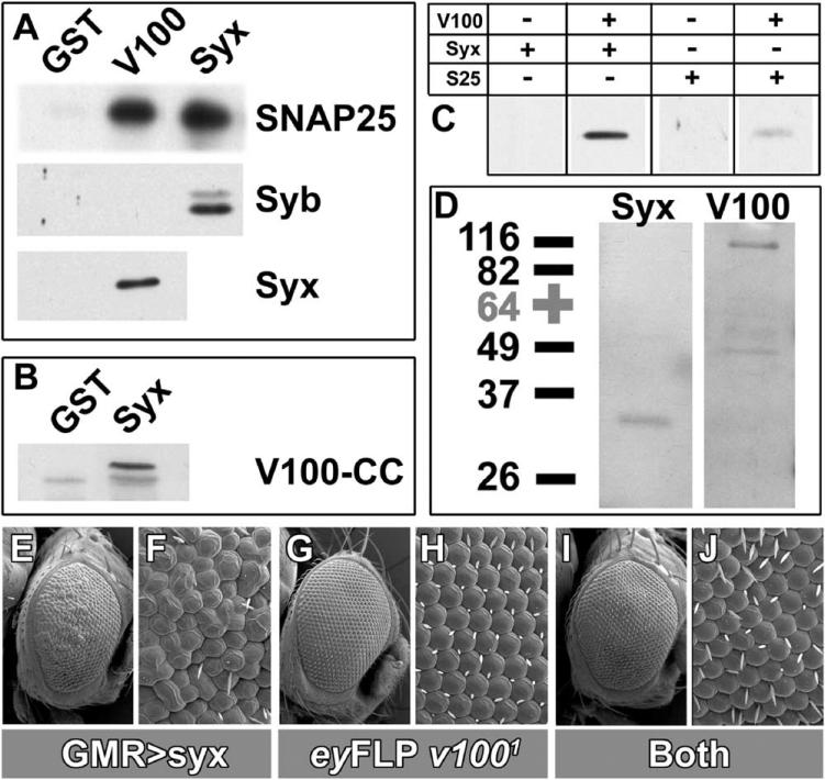
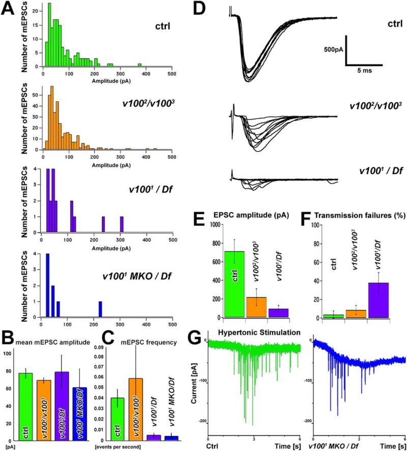
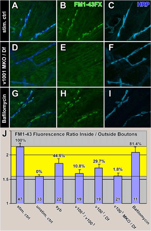
The V(0) complex forms the proteolipid pore of an ATPase that acidifies vesicles. In addition, an independent function in membrane fusion has been proposed largely based on yeast vacuolar fusion experiments. We have isolated mutations in the largest V(0) component vha100-1 in flies in an unbiased genetic screen for synaptic malfunction. The protein is only required in neurons, colocalizes with markers for synaptic vesicles as well as active zones, and interacts with t-SNAREs. Loss of vha100-1 leads to vesicle accumulation in synaptic terminals, suggesting a deficit in release. The amplitude of spontaneous release events and release with hypertonic stimulation indicate normal levels of neurotransmitter loading, yet mutant embryos display severe defects in evoked synaptic transmission and FM1-43 uptake. Our data suggest that Vha100-1 functions downstream of SNAREs in synaptic vesicle fusion.


Figures

Figure 1
Figure 1. Isolation, Identification, and Presynaptic Rescue of Mutations in v100
(A) Phototaxis of eyFLP control, v1001, v1002, and n-syb flies using the counter-current Benzer assay. (B) Electroretinogram recordings. eyFLP v100 mutants lack “on” and “off” transients (red circles), indicative of a failure to evoke a postsynaptic response. Photoreceptor-specific expression of v100 in an eyFLP v100 mutant background restores both “on” and “off.” (C) Twenty-seven kilobyte genomic region containing the P element found to be closest to the lethal mutations using recombination mapping (KG6567, recombination rate 1/19,364). (D) Identified point mutations of five alleles. CG1709-RC annotation shown in blue; cDNA used for the rescue experiments shown in green. (E) The protein sequence containing the point mutations of v1002 and v1003 is highly conserved. (F) Protein sequence homology of V100 with the fly subunits a2–a4 and the human, mouse, worm, and yeast subunit a1 homologs. (G) V100 predicted protein structure. The N terminus is cytosolic and contains two coiled-coil domains (gray gradients). The protein has seven or nine transmembrane domains (TMDs), depending on the transmembrane prediction algorithm. Depicted are seven TMDs; the two other possible TMDs are the loops depicted between TMD 2/3 and 4/5. The point mutations of the v100 alleles are marked 1–5. The protein sequences predicted to be deleted in the mutant splice variants is marbled. (H) In situ hybridizations of third instar larval brains (top) and stage-15 embryos (bottom). Antisense staining on the left shows strong nervous system expression. The sense control is shown on the right.
Figure 2
Figure 2. Drosophila V100 Rescues Vesicle Trafficking, but Not an Acidification Defect in Yeast
(A) Growth of ΔVPH1,ΔSTV1 double-mutant yeast expressing a mock control vector, yeast VPH1, or fly v100 at pH 5.5 and pH 7.5. Qualitative growth evaluation on the left; original plates on the right. Note that only rescue with VPH1 allows normal growth at pH 7.5. (B) FM4-64 pulse chase experiments with the yeast strains described in (A). Depicted is the ratio of fluorescence intensity on the vacuole compared to fluorescence intensity on the cell membrane. Note that all mutants exhibit translocation of FM4-64 to the vacuole, however with significantly different kinetics. Error bars are SEM. (C–H) 3D-Hightfield visualization of the 5 min and 90 min time points for all three genotypes in the experiment performed at pH 5.5 quantified in (B). Fluorescence intensity is visualized as relief and color-coded using the color map shown in (C). FM4-64 fluorescence on the outer membrane is marked between arrows; intracellular fluorescence on the vacuole is marked between arrowheads.
Figure 3
Figure 3. Ultrastructural Analysis of Mutant Photoreceptor Terminals in the Lamina
(A) 3D visualization of control photoreceptor neurons stained with mAb 24B10. la: lamina; me: medulla. (B) Visualization of v1001 mutant photoreceptor neurons as in (A) projecting into a heterozygous control brain. (C) Cross-section through a control lamina, showing the hexagonal pattern of cartridges, synaptic units that are demarcated by epithelial glia (blue) and contain the presynaptic photoreceptor terminals (green). (D) Lamina cross-section containing v1001 mutant photoreceptor terminals. The overall cartridge structure is unaltered. Scale bar in (D) for (C)–(D): 2 μm. (E–G) TEM micrographs of cartridges containing control, v1001, and n-syb mutant terminals, respectively. Demarcating glia are colored blue and photoreceptor terminals green to accentuate the structures. Scale bar in (E) for (E)–(G): 1 μm. (H–J) Individual terminals that are boxed in (E)–(G). Arrows: active zones; arrowheads: capitate projections (glial invaginations); double arrowheads: mitochondria. Scale bar in (H) for (H)–(J): 500 nm. (K–N) Quantification of organelles. Both v1001 and n-syb mutant terminals exhibit an increase in vesicle density of about 2.5-fold and a reduced number of capitate projections. Error bars are SEM.
Figure 4
Figure 4. Synaptic Localization of V100
(A) Adult whole-mount brain 3D visualization. V100 staining in the neuropils and cell bodies is shown in magenta. Photoreceptor-specific staining with mAb 24B10 is shown in green. The laminae are cut open on both sides revealing cross-sections of these synaptic structures. (B–D) High-resolution confocal scans of lamina cross-sections. Note the donut-like shape of V100 (magenta) and Syt (green) staining, characteristic of the localization of presynaptic photoreceptor terminals in cartridges (compare TEM of cartridges in Figure 3). Scale bar: 5 μm. (E and F) Fillet of a stage-17 embryo exposing the CNS (top left) and synapses on the body wall musculature. Green: Syx; red: V100; blue: HRP. The V100 channel is separately shown in (F). Arrows: NMJs at muscle 6/7. Scale bar: 50 μm. (G–J) High-resolution confocal scan of a control embryonic NMJ on muscle 4. DLG (blue) demarcates the NMJ border, whereas V100 (red) and n-Syb (green) colocalize presynaptically. The V100 channel is shown separately in (J). (K–N) High-resolution confocal scan of a v1001/Df embryonic NMJ on muscle 4. The staining is identical to that in (G)–(J) except that V100 staining is reduced to background levels. The V100 channel is shown separately in (N). (O–R) High-resolution 3D-deconvolved confocal sections of muscle 6/7 boutons at the third instar larval NMJ. Green: active zones labeled with mAb nc82; red: V100; blue: vesicles labeled with anti-Syt. Most V100 staining colocalizes with Syt but also extends beyond Syt at active zones (arrowheads). Scale bar: 1 μm. (S–W) Boxed region of (O) shown without pixel interpolation at original pixel size of 90 × 90 nm. (S) and (T) are three-channel composites before and after 3D deconvolution. The three channels are shown separately in (U)–(W). (X) Quantification of V100 and Syt fluorescence in five independent 3D-deconvolved datasets in threshold-segmented nc82-stained domains (shown as percentage of nc82 staining overlap). Note that V100 exhibits 50% more colocalization with the active zone marker nc82 than Syt. Error bars are SEM.
Figure 5
Figure 5. V100 Interacts with SNAREs
(A) GST pull-down assays. Top: GST (negative control), GST-V100, and GST-Syx. SNAP-25, Syb, and Syx indicate His-tagged recombinant proteins as well as the antibodies used to probe the Western blots. All fusion proteins are lacking transmembrane domains. (B) GST pull-down assay using GST-Syx to pull down a His-tagged N-terminal domain containing the two coiled-coils of V100 (V100-CC). (C) Coimmuoprecipitation (CoIP) experiments using anti-V100 coated beads. Addition of mixtures of recombinant V100 and Syx or V100 and SNAP-25, but not Syx or SNAP-25 alone, can be immunoprecipitated. (D) CoIP experiment from fly head extract using anti-Syx-coated beads. Syx as well as native full-length V100 are immunoprecipitated. (E and F) Scanning electron microscopic (SEM) images of a fly eye with photoreceptor-specific (GMR-Gal4) expression of Syx (genotype: eyFLP/+; GMR-Gal4, UAS-Syx/+; FRT cl/TM3, Sb). Overexpression of Syx is toxic and causes developmental defects resulting in a rough-eye phenotype. (G and H) SEM images of an eyFLP v1001 fly eye showing wild-type morphology (genotype: eyFLP/+; +/CyO; FRT cl/FRT v1001). (I and J) SEM images of an eyFLP v1001 fly eye with photoreceptor-specific Syx expression show suppression of the Syx overexpression toxicity (genotype: eyFLP/+; GMR-Gal4, UAS-Syx/+; FRT cl/FRT v1001).
Figure 6
Figure 6. Electrophysiological Analysis of v100 Alleles at the Embryonic NMJ
(A) Distribution of miniature excitatory postsynaptic currents amplitudes in control (green), v1002/v1003 (orange), v1001/Df (violet), and v1001 MKO/Df (blue). Note that the number of events analyzed is much lower in v1001/Df and v1001 MKO/Df because mEPSCs are rare. (B) The mean amplitude of mEPSCs in all four genotypes shows no significant difference. ctrl: 77.48 ± 5.34 (n = 161), v1002/v1003 69.15 ± 3.19 (n = 330), v1001/Df 79.29 ± 19.23 (n = 16), v1001 MKO/Df 60.93 ± 23.91 (n = 7). All error bars are SEM. (C) The frequency of mEPSCs is strongly reduced in v1001/Df and v1001 MKO/Df compared to control. v1002/v1003 NMJs display an mEPSC frequency that is on average not significantly different from control but show higher variability (note size of error bars). ctrl 0.041 ± 0.009 (n = 15), v1002/v1003 0.060 ± 0.035 (n = 13), v1001/Df 0.005 ± 0.002 (n = 6), v1001 MKO/Df 0.004 ± 0.003 (n = 6). All error bars are SEM. (D) Representative evoked responses (EPSCs) at the embryonic NMJ. (E) EPSC amplitude is significantly reduced in both v1002/v1003 and v1001/Df. (F) v1001/Df but not v1002/v1003 show a significant number of transmission failures upon stimulation. (G) Hypertonic stimulation using a 3 s application of 850 mM sucrose. Shown are control (green) and the strongest allelic combination v1001 MKO/Df (blue). Hypertonic release exists in all genotypes, albeit at a reduced level in the mutants. The mean amplitude of release events is not significantly different between control and mutant (see Figure S4).
Figure 7
Figure 7. FM1-43 Uptake at the Embryonic NMJ
(A–C) FM1-43FX uptake experiment using a 5 min 30 mM K+ stimulation paradigm. (D–F) FM1-43FX uptake using the same stimulation paradigm in a v1001 MKO/Df mutant. (G–I) FM1-43FX uptake using the same stimulation paradigm after 15 min preincubation with 1 μM Bafilomycin A1 (Baf) as well as 1 μM Baf in the FM1-43 staining solution. (J) Quantification of FM1-43 uptake experiments in stimulated control (A–C), unstimulated control (Figures S4C–S4E), n-syb (Figures S4F–S4H), v1002/v1003 (Figures S4I–S4K), v1001/Df (Figures S4L–S4N), v1001 MKO/Df (D–F), and control with added Baf (G–I). Shown is the ratio of the mean fluorescence intensity of the FM1-43 channel within the boundaries of HRP staining versus muscle background in 3D high-resolution confocal datasets. The yellow area marks the boundaries defined by unstimulated and stimulated control. Percentages of FM1-43 uptake are given with respect to these boundaries. The number of individual embryos analyzed is indicated inside bars. Error bars are SEM.

Comment in

  • A new view of an old pore.
    Bajjalieh S. Bajjalieh S. Cell. 2005 May 20;121(4):496-497. doi: 10.1016/j.cell.2005.05.002. Cell. 2005. PMID: 15907459

References

    1. Amara SG, Kuhar MJ. Neurotransmitter transporters: recent progress. Annu. Rev. Neurosci. 1993;16:73–93. - PubMed
    1. Aravamudan B, Fergestad T, Davis WS, Rodesch CK, Broadie K. Drosophila UNC-13 is essential for synaptic transmission. Nat. Neurosci. 1999;2:965–971. - PubMed
    1. Bayer MJ, Reese C, Bühler S, Peters C, Mayer A. Vacuole membrane fusion: V0 functions after trans-SNARE pairing and is coupled to the Ca2+-releasing channel. J. Cell Biol. 2003;162:211–222. - PMC - PubMed
    1. Bennett MK, Calakos N, Kreiner T, Scheller RH. Synaptic vesicle membrane proteins interact to form a multimeric complex. J. Cell Biol. 1992;116:761–775. - PMC - PubMed
    1. Benzer S. Behavioral mutants of Drosophila isolated by countercurrent distribution. Proc. Natl. Acad. Sci. USA. 1967;58:1112–1119. - PMC - PubMed

Publication types

MeSH terms

LinkOut - more resources