. 2005 Jul 8;332(3):670-6.
doi: 10.1016/j.bbrc.2005.05.006.

Putative tumor suppressor RASSF1 interactive protein and cell death inducer C19ORF5 is a DNA binding protein

Affiliations

Putative tumor suppressor RASSF1 interactive protein and cell death inducer C19ORF5 is a DNA binding protein

Leyuan Liu et al. Biochem Biophys Res Commun. .

Abstract

C19ORF5 is a homologue of microtubule-associated protein MAP1B that interacts with natural paclitaxel-like microtubule stabilizer and candidate tumor suppressor RASSF1A. Although normally distributed throughout the cytosol, C19ORF5 specifically associates with microtubules stabilized by paclitaxel or RASSF1A. At sufficiently high concentrations, C19ORF5 causes mitochondrial aggregation and genome destruction (MAGD). The accumulation on hyperstabilized microtubules coupled to MAGD has been proposed to mediate tumor suppression by the taxoid drug family and RASSF1A. Here, we show that the C-terminus of C19ORF5 (C19ORF5C) interacts with mitochondria-associated DNA binding protein, LRPPRC, in liver cells. Like LRPPRC, C19ORF5 also binds DNA with an affinity and specificity sufficient to be of utility in DNA affinity chromatography to purify homogeneous recombinant C19ORF5C from bacterial extracts. Homogeneous C19ORF5 exhibited no intrinsic DNase activity. Deletion mutagenesis indicated that C19ORF5 selectively binds double stranded DNA through its microtubule binding domain. These results suggest C19ORF5 as a DNA binding protein similar to microtubule-associated proteins tau and MAP2.


Figures

Fig. 1
Fig. 1
Interaction of C19ORF5 with DNA binding protein LRPPRC. (A) Interaction of recombinant C19ORF5 and LRPPRC in mammalian extracts. GFP-LRPPRC [1] from cell extracts was captured by purified GST-C19ORF5 immobilized on GSH beads and then visualized by immunoblot with anti-GFP antibody in assays described under Materials and methods. Cells were transfected with GFP-LRPPRC or GFP alone as indicated, extracts containing about 200 μg of total protein were loaded directly (lanes 2–4), or incubated with beads loaded with 20 lg of purified GST-C19ORF5 or GST prior to analysis as indicated (lanes 5–8). Content of the agarose beads was analyzed in lanes 5–8. Lane 1, protein standards. (B) Co-immunoprecipitation of native LRPPRC and C19ORF5 from HepG2 cells. C19ORF5 was immunoprecipitated (IP) with anti-C19ORF5 (monoclonal 4G1) from lysates of HepG2 cells and then analyzed on immunoblot (IB) with 4G1 or anti-LRPPRC (monoclonal 4C12). The indicated blots represent the lysate from 2.5 × 10⁵ cells. FL, full-length C19ORF5 predicted by translation; SC, 56 kDa short chain of C19ORF5; and Ab, mouse IgG heavy chain.
Fig. 2
Fig. 2
Purification of GST-C19ORF5C to homogeneity by combined GSH and DNA affinity chromatography. GST-C19ORF5 (residues 667–1059) was expressed in bacteria, extracted, and subjected to affinity chromatography on GSH or DNA immobilized to agarose beads as described under Materials and methods. Fractions were analyzed by SDS–PAGE and the protein was visualized by Coomassie blue stain. Lane 1, protein standards with the indicated apparent molecular mass; lane 2, GSH eluate from GSH beads of extract from bacteria expressing only the fusion tag GST; lane 3, whole extract of cells expressing GST-C19ORF5; lane 4, GSH eluate of the extract in lane 3 from GSH beads; lane 5, GSH eluate of the extract from lane 4 captured on DNA–agarose beads; lane 6, 0.5 M NaCl eluate from DNA–agarose beads; lane 7, repurification of the eluate from lane 6 on GSH beads; and lane 8, residual DNA–agarose beads from lane 6 after the 0.50 M NaCl elution. About 10 μg protein was applied to lane 3.
Fig. 3
Fig. 3
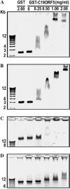
Interaction of purified GST-C19ORF5C with different types of DNA. (A) Double stranded, supercoiled pBluescript SK plasmid, (B) double stranded BamHI linearized pBluescript SK, (C) double stranded HepG2 genomic DNA (10 μg/ml), and (D) single strand ϕ X174 virion DNA were incubated with the indicated amounts of homogeneous GST-C19ORF5 or GST for 1 h at 37 °C.
Fig. 4
Fig. 4
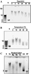
Influence of time, temperature, divalent cations, and protein cross-linking on association of purified GST-C19ORF5C with DNA. (A) Linearized pBluescript SK plasmid (10 μg/ml) was incubated with GST-C19ORF5 (2 mg/ml) for the indicated times at 37 °C or (B) the indicated temperature for 1 h. N, no GST-C19ORF5 added. (C) Effect of divalent cations (10 mM) and glutaraldehyde-induced protein cross-linking (GA) on DNA mobility. Assays contained 10 μg/ml of linearized pBluescript SK, 2 mg/ml GST-C19ORF5 protein for 1 h at 37 °C. No, GST-C19ORF5 with no divalent cations added; NP, a reaction mixture depleted of protein with the QLAquick PCR purification kit for extraction of DNA.
Fig. 5
Fig. 5
Identification of the DNA binding domain of C19ORF5. (A) Sequence domain structure of C19ORF5. Full-length C19ORF5 and the 393 amino acid residue C19ORF5C (D667–F1059) with residues flanking constructs utilized in this study are indicated. Assignment of the minimum DNA and microtubule binding domain is based on an estimate of the minimum length of a truncated product of GST-A867–E966 that bound microtubules and DNA, and homology with the microtubule binding domain of MAP1A and MAP1B [4]. F967–A991 is the MAGD domain. (B) Recombinant GST-tagged C19ORF5 subdomains expressed in bacteria. GST, D667–S766, and S767–L866 were purified by GSH affinity. A867–E966, S767–E966, and D667–F1059 were purified by both GSH and DNA affinity. Insoluble F967–F1059 was extracted directly from cells with SDS buffer. Each lane was loaded with about 10 μg protein that was visualized with Coomassie blue. (C) Specificity of monoclonal antibody 4G1. About 40 μg of the indicated expression products was analyzed by SDS–PAGE and stained with Coomassie blue. About 400 ng was subjected to immunoblot with monoclonal antibody 4G1 as described under Materials and methods. (D) DNA binding of C19ORF4 subdomains indicated by gel shift assay. The indicated purified expression products (about 2 mg/ml) were added to 10 μg/ml of 3 kb linear pBluescript SK DNA and incubated for 1 h at 37 °C prior to analysis.

Similar articles

Cited by

References

    1. Liu L, Vo A, Liu G, McKeehan WL. Novel complex integrating mitochondria and the microtubular cytoskeleton with chromosome remodeling and tumor suppressor RASSF1 deduced by in silico homology analysis, interaction cloning in yeast, and colocalization in cultured cells. In Vitro Cell. Dev. Biol. Anim. 2002;38:582–594. - PMC - PubMed
    1. Dallol A, Agathanggelou A, Fenton SL, Ahmed-Choudhury J, Hesson L, Vos MD, Clark GJ, Downward J, Maher ER, Latif F. RASSF1A interacts with microtubule-associated proteins and modulates microtubule dynamics. Cancer Res. 2004;64:4112–4116. - PubMed
    1. Liu L, Vo A, McKeehan WL. Specificity of the methylation-suppressed A isoform of candidate tumor suppressor RASSF1 for microtubule hyperstabilization is determined by cell death inducer C19ORF5. Cancer Res. 2005;65:1830–1838. - PubMed
    1. Liu L, Vo A, Liu G, McKeehan WL. Distinct structural domains within C19ORF5 support association with stabilized microtubules and mitochondrial aggregation and genome destruction. Cancer Res. 2005;65:4191–4201. - PMC - PubMed
    1. Song MS, Chang JS, Song SJ, Yang TH, Lee H, Lim DS. The centrosomal protein RABP1 regulates mitotic progression by recruiting RASSF1A to spindle poles. J. Biol. Chem. 2005;280:3920–3927. - PubMed

Publication types

MeSH terms