. 2005 Jun;73(6):3210-8.
doi: 10.1128/IAI.73.6.3210-3218.2005.

Role of sialic acid and complex carbohydrate biosynthesis in biofilm formation by nontypeable Haemophilus influenzae in the chinchilla middle ear

Affiliations

Role of sialic acid and complex carbohydrate biosynthesis in biofilm formation by nontypeable Haemophilus influenzae in the chinchilla middle ear

Joseph Jurcisek et al. Infect Immun. 2005 Jun.

Abstract

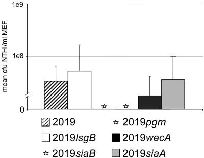
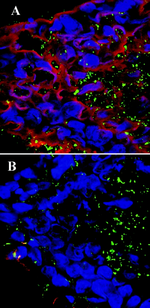
Nontypeable Haemophilus influenzae (NTHI) is an important pathogen in respiratory tract infections, including otitis media (OM). NTHI forms biofilms in vitro as well as in the chinchilla middle ear, suggesting that biofilm formation in vivo might play an important role in the pathogenesis and chronicity of OM. We've previously shown that SiaA, SiaB, and WecA are involved in biofilm production by NTHI in vitro. To investigate whether these gene products were also involved in biofilm production in vivo, NTHI strain 2019 and five isogenic mutants with deletions in genes involved in carbohydrate biosynthesis were inoculated into the middle ears of chinchillas. The wild-type strain formed a large, well-organized, and viable biofilm; however, the wecA, lsgB, siaA, pgm, and siaB mutants were either unable to form biofilms or formed biofilms of markedly reduced mass, organization, and viability. Despite their compromised ability to form a biofilm in vivo, wecA, lsgB, and siaA mutants survived in the chinchilla, inducing culture-positive middle ear effusions, whereas pgm and siaB mutants were extremely sensitive to the bactericidal activity of chinchilla serum and thus did not survive. Lectin analysis indicated that sialic acid was an important component of the NTHI 2019 biofilm produced in vivo. Our data suggested that genes involved in carbohydrate biosynthesis and assembly play an important role in the ability of NTHI to form a biofilm in vivo. Collectively, we found that when modeled in a mammalian host, whereas biofilm formation was not essential for survivability of NTHI in vivo, lipooligosaccharide sialylation was indispensable.


Figures

FIG. 1.
FIG. 1.
Gross whole-mount images of bullae recovered 5 days postchallenge from chinchillas inoculated transbullarly with NTHI strain 2019 or a mutant derivative thereof. Brackets indicate biofilms (if present), and TM indicates tympanic membrane (or ear drum). (A) Bulla recovered from a naive chinchilla for comparison. Panel B shows large biofilm formed by strain 2019 in the chinchilla middle ear cavity 5 days after challenge. Strains 2019lsgB and 2019siaA (panels C and D, respectively) induced the formation of very small biofilms, easily identifiable only by stereomicroscopy. Strain 2019wecA formed a biomass in only two of the six challenged middle ears (panel E). In the remaining four middle ears, despite the presence of marked erythema, there were no discernible biomasses (panel F). Strains 2019siaB and 2019pgm (panels G and H, respectively) did not induce the formation of a biofilm that could be identified upon gross examination. Magnification all panels, 5×.
FIG. 2.
FIG. 2.
(A) H&E stain of an OCT-embedded biofilm produced by strain 2019 in the chinchilla middle ear (5× magnification). Inset (100× magnification) clearly demonstrates what appears to be numerous water channels present within the biofilm. (B) TEM analysis of OCT-embedded biofilm formed by NTHI strain 2019 in the middle ear of a chinchilla. Arrows indicate bacteria surrounded by the biofilm matrix. This section was incubated with Sambucus nigra lectin conjugated to 15-nm gold beads. Note that the gold beads are seen bound to the biofilm matrix and not to the bacteria within the biofilm.
FIG. 3.
FIG. 3.
Mean CFU NTHI per ml middle ear fluid (± standard deviations) as recovered from each of six middle ears challenged with either strain 2019 or one of its five deletion mutants. Strains 2019siaB and 2019pgm did not yield culture positive effusions in any of the six challenged middle ears (see asterisks in place of bars in figure).
FIG. 4.
FIG. 4.
Confocal microscopy images of whole-mount sections of chinchilla inferior bulla (white arrows indicate bone of the inferior bulla in each panel) stained with LIVE/DEAD bacterial stain 5 days after direct challenge with NTHI. The parent strain, 2019 (panel A) induced the formation of a large biofilm (white bracket) with long finger-like projections that appear to be separated by water channels that do not contain bacteria. These projections extend well into the middle ear space. Strain 2019pgm (panel B) formed a very small biofilm with what also appears to be water channels; however, this biofilm appeared to be less organized. Strains 2019lsgB (panel C) and 2019siaA (panel D) formed very dense biofilms with no apparent water channels. A much larger population of dead NTHI (red stain, panel C) was observed in the biofilm induced by strain 2019lsgB than seen in that produced by the parental isolate at this time point postchallenge of themiddle ear. Strain 2019wecA induced the formation of a biomass in only two of the six challenged middle ears (panel E). These biomasses lacked architecture and were predominantly nonviable (bone of inferior bulla was viable; however, it appears black in this image because it was below the plane that was optimal for imaging the fluorescent bacterial biomass). In the majority of middle ears challenged with 2019wecA, there were no detectable biofilms; however, there were focal areas of bright fluorescence on the mucosal surface and the bone of the inferior bulla appears to be considerably thickened (panel F), suggesting the presence of viable bacteria and demonstrating that an inflammatory response, with new bone formation, had occurred following challenge of the depicted middle ear with strain 2019wecA. Strain 2019siaB (panel G) did not form detectable biofilms in the chinchilla middle ear 5 days after direct inoculation. A similarly stained naïve bulla is presented in panel H to demonstrate the normal thickness of the bone of the inferior bulla in an unchallenged chinchilla middle ear.
FIG. 5.
FIG. 5.
SEM images of parent strain 2019 (panel A) and strain 2019wecA (panel B) incubated for 3 days on primary human bronchus cells. The parent strain formed organized microcolonies of bacteria (see arrow in panel A), whereas the mutant strain was seen to grow primarily as a monolayer of individual bacteria on the surface of these mucosal epithelial cells (panel B). The magnification of each panel is 6,000×.
FIG. 6.
FIG. 6.
Composite of confocal images obtained following incubation of an OCT-embedded biofilm produced by strain 2019 in the chinchilla middle ear with two fluorochrome-conjugated lectins. (A) Binding of the lectin Sambucus nigra (SNA-TRITC) is shown in red. The specificity of this lectin is for sialic acid α-2-6 galactose, and in these images it is shown binding to the biofilm matrix. Macchia amurensia lectin (MAA-FITC in green), which has specificity for sialic acid α-2-3 galactose and lactose, bound to the LOS of the NTHI present within the biofilm. (B) Lectin labeling obtained after neuraminidase treatment of a serial section of the same OCT-embedded biofilm that is shown in panel A. Neuraminidase removed labeling by SNA-TRITC of the biofilm completely, confirming the presence of sialic acid in an α-2-6 linkage within the biofilm matrix (4). There was minimal change observed in the binding of MAA-FITC to NTHI within the biofilm after neuraminidase treatment. In addition, these sections show the infiltration of the biofilm by inflammatory cells (the nuclei of which are labeled blue with a DNA stain [To-Pro3]). The magnification of each panel is 1,200×.

Similar articles

Cited by

References

    1. Andreoni, J., H. Kayhty, and P. Densen. 1993. Vaccination and the role of capsular polysaccharide antibody in prevention of recurrent meningococcal disease in late complement component-deficient individuals. J. Infect. Dis. 168:227-231. - PubMed
    1. Bakaletz, L. O., B. D. Baker, J. A. Jurcisek, H. Harrison, L. A. Novotny, J. E. Bookwalter, R. Mungur, and R. S. Munson, Jr. Demonstration of type IV pilus expression and a twitching phenotype by Haemophilus influenzae. Infect. Immun., in press. - PMC - PubMed
    1. Bakaletz, L. O., B. J. Kennedy, L. A. Novotny, G. Duquesne, J. Cohen, and Y. Lobet. 1999. Protection against development of otitis media induced by nontypeable Haemophilus influenzae by both active and passive immunization in a chinchilla model of virus-bacterium superinfection. Infect. Immun. 67:2746-2762. - PMC - PubMed
    1. Brinkman-Van der Linden, E. C., J. L. Sonnenburg, and A. Varki. 2002. Effects of sialic acid substitutions on recognition by Sambucus nigra agglutinin and Maackia amurensis hemagglutinin. Anal. Biochem. 303:98-104. - PubMed
    1. Campagnari, A. A., M. R. Gupta, K. C. Dudas, T. F. Murphy, and M. A. Apicella. 1987. Antigenic diversity of lipooligosaccharides of nontypable Haemophilus influenzae. Infect. Immun. 55:882-887. - PMC - PubMed

Publication types

Substances

LinkOut - more resources