Comparative Study
. 2005 Jun;73(6):3228-41.
doi: 10.1128/IAI.73.6.3228-3241.2005.

Comparison of Salmonella enterica serovar Typhimurium colitis in germfree mice and mice pretreated with streptomycin

Affiliations
Comparative Study

Comparison of Salmonella enterica serovar Typhimurium colitis in germfree mice and mice pretreated with streptomycin

Bärbel Stecher et al. Infect Immun. 2005 Jun.

Abstract

Salmonella enterica subspecies 1 serovar Typhimurium is a common cause of bacterial enterocolitis. Mice are generally protected from Salmonella serovar Typhimurium colonization and enterocolitis by their resident intestinal microflora. This phenomenon is called "colonization resistance" (CR). Two murine Salmonella serovar Typhimurium infection models are based on the neutralization of CR: (i) in specific-pathogen-free mice pretreated with streptomycin (StrSPF mice) antibiotics disrupt the intestinal microflora; and (ii) germfree (GF) mice are raised without any intestinal microflora, but their intestines show distinct physiologic and immunologic characteristics. It has been unclear whether the same pathogenetic mechanisms trigger Salmonella serovar Typhimurium colitis in GF and StrSPF mice. In this study, we compared the two colitis models. In both of the models Salmonella serovar Typhimurium efficiently colonized the large intestine and triggered cecum and colon inflammation starting 8 h postinfection. The type III secretion system encoded in Salmonella pathogenicity island 1 was essential in both disease models. Thus, Salmonella serovar Typhimurium colitis is triggered by similar pathogenetic mechanisms in StrSPF and GF mice. This is remarkable considering the distinct physiological properties of the GF mouse gut. One obvious difference was more pronounced damage and reduced regenerative response of the cecal epithelium in GF mice. Overall, StrSPF mice and GF mice provide similar but not identical models for Salmonella serovar Typhimurium colitis.


Figures

FIG. 1.
FIG. 1.
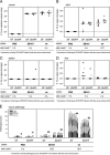
Preliminary time course study of Salmonella serovar Typhimurium infection in nonpretreated SPF, StrSPF, and GF mice. Seven nonpretreated SPF mice, six StrSPF mice, and five GF mice were infected with 5 × 10⁷ CFU of wild-type SL1344 intragastrically. At 4 h, 8 h, and 20 h p.i. one, two, or three animals were sacrificed, as indicated in panel A. (A) Bacterial loads in the cecal contents. (B) Histopathological analyses: H&E-stained sections of cryo-embedded cecal tissue were scored with respect to edema in the submucosa, PMN infiltration (PMN inf.), reduction in the number of goblet cells, and desquamation, erosion, and ulceration of the epithelial layer (see Materials and Methods). Scores are plotted as stacked vertical bars. The dotted line indicates the limit of detection (cecal contents).
FIG. 2.
FIG. 2.
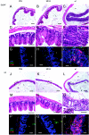
Pathological changes in ceca of nonpretreated SPF, StrSPF, and GF mice infected with Salmonella serovar Typhimurium. Thin sections (5 μm) of cryo-embedded cecal tissues of mice from the experiment described in the legend to Fig. 1 were stained with H&E as described in Materials and Methods. Nonpretreated SPF mice (A and D), StrSPF mice (B, E, and G), and GF mice (C, F, and H) were infected with wild-type strain SL1344 for 20 h. Panels D, E, and F are enlargements of the sections indicated by the boxes in panels A, B, and C, respectively. Panels G and H are enlargements of the sections indicated by the boxes in panels E and F, respectively. The arrows in panels G and H indicate PMN. e, submucosal edema; L, intestinal lumen; er, erosion. (A, B, and C) Scale bars = 200 μm. (D, E, and F) Scale bars = 100 μm. (G and H) Scale bars = 20 μm.
FIG.3.
FIG.3.
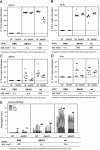
Infection of GF and StrSPF mice with Salmonella serovar Typhimurium wild-type strain and strain SB161 for 24 h. Groups of five StrSPF mice or GF mice were infected intragastrically for 24 h with 5 × 10⁷ CFU of serovar Typhimurium strain SL1344 (wild type) or SB161 or were mock infected with PBS (A to D) (solid circles). In an independent experiment, groups of two StrSPF mice or two GF mice were infectedintragastrically for 24 h with 5 × 10⁷ CFU of serovar Typhimurium strain SL1344 (wild type) or SB161. One StrSPF mouse and one GF mouse were mock infected with PBS. Tissue colonization was determined, and cecal tissues were fixed in 4% formalin and embedded in paraffin. The latter mice are indicated by open circles in panels A to D and by plus signs in panel E. (A to D) Bacterial loads in the cecal contents (A), in the mLN (B), in the spleen (C), and in the liver (D). (E) Histopathological analyses. H&E-stained sections of cryo-embedded (not labeled) or paraffin-embedded (plus signs) cecal tissues were scored with respect to edema in the submucosa, PMN infiltration (PMN inf.), reduction in the number of goblet cells, and desquamation, erosion, and ulceration of the epithelial layer (see Materials and Methods). The dotted lines indicate the limit of detection. The solid lines indicate the median. n.s., not statistically significant (P ≥ 0.05); S. Tm, Salmonella serovar Typhimurium; stat. anal., statistical analysis; wt, wild type.
FIG. 4.
FIG. 4.
Histopathological analysis, infiltration of CD18+ cells, and expression of ICAM-1 in GF and StrSPF mice at 24 h p.i. Thin sections of cecal tissues of StrSPF mice (A to I) and GF mice (J to R) from the experiment described in the legend to Fig. 2 were stained as described in Materials and Methods. (A to F and J to O) H&E-stained thin sections of paraffin-embedded tissues (see Fig. 3E). Mice were mock infected with PBS (A, D, J, and M) or infected with SB161 (B, E, K, and N) or wild-type strain SL1344 (C, F, L, and O). Panels D to F and M to O are enlargements of the areas indicated by the boxes in panels A to C and J to L, respectively. The boxes in panels F and O show PMN (arrows). (G to I and P to R) Expression of ICAM-1 and infiltration of CD18+ cells. Thin sections (7 μm) of cryo-embedded tissues were stained with DAPI, rat anti-mouse CD18, hamster anti-mouse ICAM-1, and polyclonal anti-rat IgG-Cy5 and anti-hamster IgG-Cy3 antibodies as described in Materials and Methods (DNA, blue; ICAM-1, red; CD18, green). Mice were mock infected with PBS (G and P) or were infected with SB161 (H and Q) or wild-type strain SL1344 (I and R). L, intestinal lumen; e, edema; er, erosion. The arrows indicate PMN. (A to C and J to L) Scales bars = 200 μm. (D to I and M to R) Scale bars = 50 μm.
FIG.5.
FIG.5.
Infection of GF and StrSPF mice with wild-type Salmonella serovar Typhimurium and strain SB161 for 48 h. Groups of five StrSPF mice or seven or eight GF mice were infected intragastrically for 48 h with 5 × 10⁷ CFU of serovar Typhimurium strain SL1344 (wild type) or SB161 or were mock infected with PBS. In an independent experiment, groups of two StrSPF mice or two GF mice were infected intragastrically for 48 h with 5 × 10⁷ CFU of serovar Typhimurium strain SL1344 (wild type) or SB161. One StrSPF mouse or one GF mouse was mock infected with PBS. Tissue colonization was determined, and cecal tissues were fixed in 4% formalin and embedded in paraffin. These mice are represented by open circles (A to D) or by plus signs (E). We analyzed bacterial loads in the cecal contents (A), in the mLN (B), in the spleen (C), and in the liver (D) and performed histopathological analyses (E). H&E-stained sections of cryo-embedded (not labeled) or paraffin-embedded (plus signs) cecal tissues were scored with respect to edema in the submucosa, PMN infiltration (PMN inf.), reduction in the number of goblet cells, and desquamation, erosion, and ulceration of the epithelial layer (see Materials and Methods). Scores are plotted as stacked vertical bars. The dotted lines indicate the limit of detection. The solid lines indicate the median. The dagger and the asterisk indicate animals analyzed in Fig. 6M and N. n.s., not statistically significant (P ≥ 0.05); S. Tm, Salmonella serovar Typhimurium; stat. anal., statistical analysis; wt, wild type.
FIG. 6.
FIG. 6.
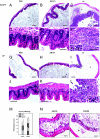
Cecal pathology at 48 h p.i. Thin sections of paraffin-embedded cecal tissues of StrSPF mice and GF mice from the experiment described in the legend to Fig. 5E (plus signs) were stained as described in Materials and Methods. Mice were mock infected with PBS (A, D, G, and J) or were infected with SB161 (B, E, H, and K) or wild-type strain SL1344 (C, F, I, and L). Panels D to F and J to L are enlargements of the areas indicated by boxes in panels A to C and G to I, respectively. The boxes in panels F and L show PMN (arrows). (M and N) Cryo-embedded tissue sections of the apical and medial part of the cecum of two wild-type SL1344-infected GF mice (indicated by a dagger and an asterisk in Fig. 5E) were analyzed. (M) Total pathological score for sections from apical and medial parts of the ceca of two mice (indicated by the asterisk and dagger, respectively). (N) H&E-stained thin section of apical and medial parts of the cecum of the same mouse (asterisk). L, intestinal lumen; e, edema; de = desquamation; PMN inf., PMN infiltration. Scale bar = 200 μm.
FIG.7.
FIG.7.
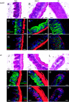
Epithelial regeneration in Salmonella serovar Typhimurium-infected GF and StrSPF mice. Serial thin sections of cryo-embedded cecal tissues of StrSPF mice (A and I) and GF mice (J and R) from the experiment described in the legend to Fig. 2 were stained as described in Materials and Methods. (A to C and J to L) H&E-stained thin sections (5 μm). Mice were mock infected with PBS (A and J) or infected with SB161 (B and K) or wild-type strain SL1344 (C and L). (D to F and M to O) Immunofluorescence analysis of the cecal epithelium. Thin sections (7 μm) were stained with DAPI, polyclonal rabbit anti-mouse cytokeratin, TRITC-phalloidin, and polyclonal anti-rabbit IgG-FITC antibodies as described in Materials and Methods. Mice were treated with PBS (D and M), SB161 (E and N), or wild-type strain SL1344 (F and O). (DNA, blue; actin, red; cytokeratin, green). (G to I and P to R) Regeneration of epithelial cells. Thin sections (7 μm) were stained with DAPI, polyclonal rabbit anti-mouse Ki-67, TRITC-phalloidin, and polyclonal anti-rabbit IgG-FITC antibodies as described in Materials and Methods. Mice were treated with PBS (G and P), SB161 (H and Q), or wild-type strain SL1344 (I and R). (DNA, blue; actin, red; Ki-67, green). L, intestinal lumen; e, submucosal edema. Scale bars = 50 μm.

References

    1. Ayabe, T., D. P. Satchell, C. L. Wilson, W. C. Parks, M. E. Selsted, and A. J. Ouellette. 2000. Secretion of microbicidal alpha-defensins by intestinal Paneth cells in response to bacteria. Nat. Immunol. 1:113-118. - PubMed
    1. Barthel, M., S. Hapfelmeier, L. Quintanilla-Martinez, M. Kremer, M. Rohde, M. Hogardt, K. Pfeffer, H. Russmann, and W. D. Hardt. 2003. Pretreatment of mice with streptomycin provides a Salmonella enterica serovar Typhimurium colitis model that allows analysis of both pathogen and host. Infect. Immun. 71:2839-2858. - PMC - PubMed
    1. Bernet, M. F., D. Brassart, J. R. Neeser, and A. L. Servin. 1994. Lactobacillus acidophilus LA 1 binds to cultured human intestinal cell lines and inhibits cell attachment and cell invasion by enterovirulent bacteria. Gut 35:483-489. - PMC - PubMed
    1. Bohnhoff, M., C. P. Miller, and W. R. Martin. 1964. Resistance of the mouse intestinal tract to experimental Salmonella infection. I. Factors which interfere with the initiation of infection by oral inoculation. J. Exp. Med. 120:805-816. - PMC - PubMed
    1. Bohnhoff, M., C. P. Miller, and W. R. Martin. 1964. Resistance of the mouse intestinal tract to experimental Salmonella infection. II. Factors responsible for its loss following streptomycin treatment. J. Exp. Med. 120:817-828. - PMC - PubMed

Publication types

LinkOut - more resources