. 2005 Jun;73(6):3271-7.
doi: 10.1128/IAI.73.6.3271-3277.2005.

Direct activation of human endothelial cells by Plasmodium falciparum-infected erythrocytes

Affiliations

Direct activation of human endothelial cells by Plasmodium falciparum-infected erythrocytes

Nicola K Viebig et al. Infect Immun. 2005 Jun.

Abstract

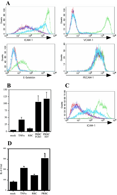
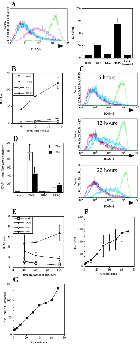
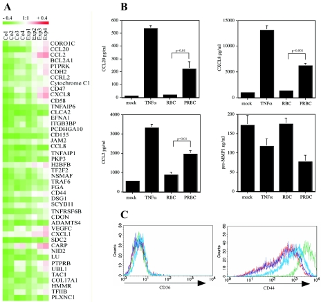
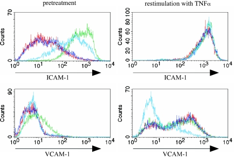
Cytoadherence of Plasmodium falciparum-infected erythrocytes (PRBC) to endothelial cells causes severe clinical disease, presumably as a of result perfusion failure and tissue hypoxia. Cytoadherence to endothelial cells is increased by endothelial cell activation, which is believed to occur in a paracrine fashion by mediators such as tumor necrosis factor alpha (TNF-alpha) released from macrophages that initially recognize PRBC. Here we provide evidence that PRBC directly stimulate human endothelial cells in the absence of macrophages, leading to increased expression of adhesion-promoting molecules, such as intercellular adhesion molecule 1. Endothelial cell stimulation by PRBC required direct physical contact for a short time (30 to 60 min) and was correlated with parasitemia. Gene expression profiling of endothelial cells stimulated by PRBC revealed increased expression levels of chemokine and adhesion molecule genes. PRBC-stimulated endothelial cells especially showed increased expression of molecules involved in parasite adhesion but failed to express molecules promoting leukocyte adhesion, such as E-selectin and vascular cell adhesion molecule 1, even after challenge with TNF-alpha. Collectively, our data suggest that stimulation of endothelial cells by PRBC may have two effects: prevention of parasite clearance through increased cytoadherence and attenuation of leukocyte binding to endothelial cells, thereby preventing deleterious immune reactivity.


Figures

FIG. 1.
FIG. 1.
P. falciparum-infected erythrocytes induce endothelial cell activation. (A) Cell surface expression levels of ICAM-1, VCAM-1, E-selectin, and PECAM-1 were determined by indirect immunostaining on HUVEC after incubation for 20 h with medium alone (red line), RBC alone (dark blue line), TNF-α (10 ng/ml) (green line), or PRBC infected with P. falciparum clone FCR3 (light blue line). (B) HUVEC were cultured in the presence of medium, RBC, TNF-α, or PRBC. Cell culture supernatants were collected after 20 h, and the IL-6 concentration was measured by the ELISA. IL-6 secretion by HUVEC treated with TNF-α (P < 0.001, compared with the medium control; n = 8) and with P. falciparum-infected erythrocytes (P < 0.001, compared with the RBC control; n = 8) was significantly increased. (C and D) Surface expression of ICAM-1 on human lung endothelial cells (C) and cell culture supernatant concentration of IL-6 (D). Human lung endothelial cells were treated as described above for panel A.
FIG. 2.
FIG. 2.
Characteristics of PRBC-mediated activation of HUVEC. (A) HUVEC were cocultured with RBC or PRBC either in direct physical contact or separated by a transwell system (pore size, 0.4 μm). Cell surface expression levels of ICAM-1 on HUVEC were detected by flow cytometry following incubation with medium (red line), RBC alone (dark blue line), TNF-α (10 ng/ml) (green line), PRBC in the transwell (light blue line), or PRBC in direct physical contact (purpleline). The IL-6 concentration was determined in cell culture supernatants by the ELISA. (B) Expression of IL-6 by HUVEC was determined after 6, 12, and 22 h of continuous incubation with medium, TNF-α, RBC, or PRBC. (C) HUVEC were incubated as described in the legend to Fig. 1A for 6, 12, and 22 h and were then analyzed for ICAM-1 surface expression levels. (D) HUVEC were incubated for 22 h as indicated in the legend to Fig. 1A. Cells were either analyzed for ICAM-1 surface expression levels or incubated in the absence of stimulators for another 18 h, after which ICAM-1 surface expression levels were determined. (E) HUVEC were incubated for different times (30 min, 60 min, and 120 min) with medium, TNF-α, RBC, or PRBC, and the IL-6 concentrations in cell culture supernatants were determined after a total incubation time of 21 h. (F and G) To study the correlation between parasite number and endothelial cell activation, HUVEC were cultured with 7 × 10⁶ RBC/ml containing PRBC at the percentages indicated, and the concentrations of IL-6 in cell culture supernatants (F) or the cell surface expression levels of ICAM-1 (G) were determined as described above.
FIG. 3.
FIG. 3.
Gene expression analysis of HUVEC after contact with PRBC. (A) RNA was isolated from RBC or PRBC-treated HUVEC and used for hybridization as described in Materials and Methods. The corresponding FDR value was 0.19417. Hierarchical clustering of genes identified by SAM as being statistically significantly differently expressed was performed using the Tiger software (version 2.2) with the average linkage clustering method (distance metric, Euclidean). (B) Results of gene expression profiling were validated at the protein level. Cell culture supernatants from HUVEC treated as described in the text were assayed for CCL20, CCL2, CXCL8, and pro-MMP-1 by specific ELISA. (C) Cell surface expression levels of CD44 and CD36 on HUVEC were determined by immunofluorescent staining and flow cytometric analysis.
FIG. 4.
FIG. 4.
Phenotype of PRBC-stimulated HUVEC after contact with TNF-α. HUVEC were incubated for 20 h with medium (red line), RBC alone (dark blue line), TNF-α (10 ng/ml) (green line), or PRBC (light blue line), and cells were washed three times with medium and cultivated for an additional 6 h with medium or with TNF-α (10 ng/ml). Cells were analyzed by flow cytometry after indirect immunostaining with antibodies recognizing ICAM-1, VCAM-1, and E-selectin. The histograms show the expression levels of the surface molecules.

References

    1. Andrews, K. T., N. K. Viebig, F. Wissing, N. Klatt, N. Oster, H. Wickert, P. Knolle, and M. Lanzer. 2003. A human schwannoma cell line supports the in vitro adhesion of Plasmodium falciparum infected erythrocytes to chondroitin-4-sulfate. Parasitol. Res. 89:188-193. - PubMed
    1. Barnwell, J. W., A. S. Asch, R. L. Nachman, M. Yamaya, M. Aikawa, and P. Ingravallo. 1989. A human 88-kD membrane glycoprotein (CD36) functions in vitro as a receptor for a cytoadherence ligand on Plasmodium falciparum-infected erythrocytes. J. Clin. Investig. 84:765-772. - PMC - PubMed
    1. Berendt, A. R., D. L. Simmons, J. Tansey, C. I. Newbold, and K. Marsh. 1989. Intercellular adhesion molecule-1 is an endothelial cell adhesion receptor for Plasmodium falciparum. Nature 341:57-59. - PubMed
    1. Clemetson, K. J., J. M. Clemetson, A. E. Proudfoot, C. A. Power, M. Baggiolini, and T. N. Wells. 2000. Functional expression of CCR1, CCR3, CCR4, and CXCR4 chemokine receptors on human platelets. Blood 96:4046-4054. - PubMed
    1. Craig, A., and A. Scherf. 2001. Molecules on the surface of the Plasmodium falciparum infected erythrocyte and their role in malaria pathogenesis and immune evasion. Mol. Biochem. Parasitol. 115:129-143. - PubMed

Publication types

Substances

LinkOut - more resources