. 2005 Jun;73(6):3422-30.
doi: 10.1128/IAI.73.6.3422-3430.2005.

Entamoeba histolytica and Entamoeba dispar utilize externalized phosphatidylserine for recognition and phagocytosis of erythrocytes

Affiliations

Entamoeba histolytica and Entamoeba dispar utilize externalized phosphatidylserine for recognition and phagocytosis of erythrocytes

Douglas R Boettner et al. Infect Immun. 2005 Jun.

Abstract

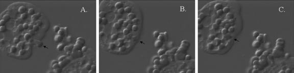
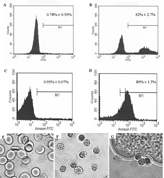
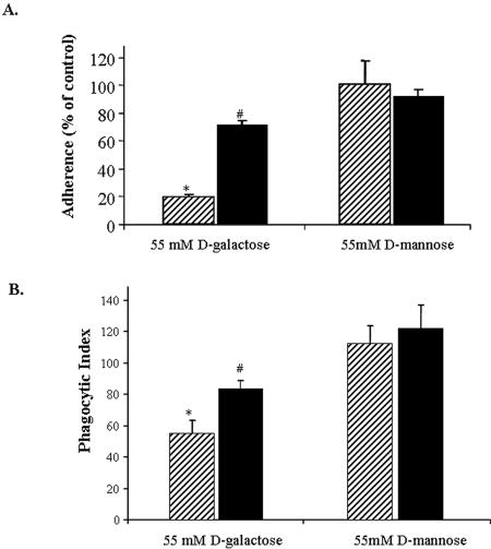
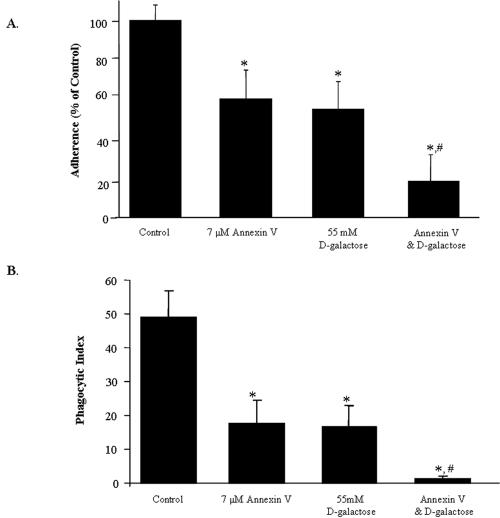
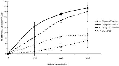
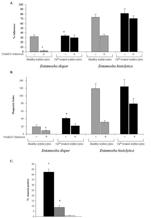
Amebic erythrophagocytosis is characteristic of invasive amebiasis, and mutants deficient in erythrocyte ingestion are avirulent. We sought to understand the molecular mechanisms underlying erythrocyte phagocytosis by Entamoeba histolytica. Following adherence to amebae, erythrocytes became round and crenulated, and phosphatidylserine (PS) was exposed on their outer membrane leaflets. These changes were similar to the effects of calcium treatment on erythrocytes, which we utilized to separate ameba-induced exposure of erythrocyte PS from the process of phagocytosis. The adherence and phagocytosis of calcium-treated erythrocytes were less inhibited by galactose than were those of healthy erythrocytes, suggesting the existence of an amebic coreceptor specific for PS. To test whether PS was recognized by amebae, calcium-treated cells were incubated with annexin V prior to adherence to or ingestion by E. histolytica. Annexin V blocked both adherence (50% +/- 12% inhibition; P < 0.05) and phagocytosis (65% +/- 10%; P < 0.05), providing evidence that at least one galactose-independent coreceptor was involved in the adherence and ingestion of red blood cells. The coreceptor was inhibited by phospho-l-serine and to a lesser extent by phospho-d-serine but not by phospho-l-threonine, which is consistent with the coreceptor functioning in the adherence and ingestion of erythrocytes via recognition of PS. We expanded our investigations to the highly related but noninvasive parasite Entamoeba dispar and demonstrated that it was deficient in red-blood-cell adherence, induction of PS exposure, and phagocytosis. These findings establish phosphatidylserine involvement in erythrophagocytosis by amebae and suggest the existence of a PS receptor on the surfaces of both E. histolytica and E. dispar.


Figures

FIG. 1.
FIG. 1.
Erythrophagocytosis by E. histolytica. An intact erythrocyte (arrow) was followed from adherence through ingestion with 40 seconds of elapsed time between each panel. (A) Entamoeba histolytica adherent to an erythrocyte with membrane distortion. (B) Erythrocyte ingested intact. (C) Erythrocyte redistributed within the ameba. A video is available online at http://www.healthsystem.virginia.edu/internet/petri-mann/movies/ehistrbc.mov.
FIG. 2.
FIG. 2.
Erythrocytes exposed to Entamoeba histolytica redistribute phosphatidylserine to the outer leaflet of the membrane and undergo morphological changes. (A to D) PS exposure was measured using annexin V-FITC staining of erythrocytes. Representative histograms of healthy erythrocytes and erythrocytes incubated with E. histolytica trophozoites are shown in panels A and B, respectively. Similarly, annexin V-FITC staining was performed on erythrocytes exposed to treatment with 2.5 mM calcium. Representative histograms of healthy erythrocytes in HEPES buffer for 48 h and erythrocytes in HEPES buffer exposed to 2.5 mM CaCl2 are shown in panels C and D, respectively. (E to G) Representative micrographs of healthy erythrocytes (panel E), crenulated erythrocytes following 48 h calcium treatment (panel F), and erythrocytes following 20 min exposure to E. histolytica (panel G).
FIG. 3.
FIG. 3.
Entamoeba histolytica cells adhere to and ingest calcium-treated erythrocytes in a relatively galactose-independent manner. (A) Adherence of E. histolytica to healthy (hatched bars) and calcium-treated (solid bars) erythrocytes. Adherent cells were defined as trophozoites with at least three adherent erythrocytes (means ± SD; n = 6; * indicates a P value of <0.001 compared to healthy cells plus d-mannose; # indicates a P value of <0.01 compared to healthy cells plus d-galactose). (B) Phagocytosis of healthy (hatched bars) or calcium-treated (solid bars) erythrocytes by amebae (reported according to the phagocytic index, i.e., the average number of erythrocytes ingested multiplied by the percentage of amebae positive for ingestion). Following calcium treatment (2.5 mM), erythrocytes were stained with TAMRA and then spun onto amebae and incubated at 37°C for 15 min at a 10:1 erythrocyte-to-ameba ratio. Unengulfed erythrocytes were lysed in water, and amebae containing ingested erythrocytes were counted by microscopy. Data are reported as means ± SD. P values were determined by a two-tailed t test for healthy and calcium-treated cells (* indicates a P value of <0.001 compared to healthy erythrocytes with d-mannose; # indicates a P value of <0.01 compared to healthy erythrocytes with d-galactose; n = 6).
FIG. 4.
FIG. 4.
Adherence and phagocytosis of calcium-treated erythrocytes are blocked by annexin V and galactose in an additive manner. (A) Human erythrocytes were preincubated for 24 h with 2.5 mM CaCl2 in order to induce apoptotic-like surface changes. These cells were washed and then incubated with 7 μM annexin V. Each of the three samples was statistically significantly different from the control (* indicates a P value of <0.05 compared to calcium-treated erythrocytes without d-galactose or annexin V; n = 10). The addition of annexin V to galactose further reduced adherence by 15% when compared to galactose alone (# indicates a P value of <0.01; n = 10). (B) Following calcium treatment (2.5 mM) cells were stained with TAMRA, quenched, and preincubated with 7 μM annexin V. Cells were spun onto amebae and incubated at 37°C for 15 min at a 5:1 erythrocyte-to-ameba ratio. Uningested erythrocytes were lysed in water, and the cells were counted by microscopy. Data are reported as means ± SD. Differences between control cells (calcium-treated cells without annexin or galactose) and all other treatments were statistically significant (* indicates a P value of <0.0001 compared to the control). Also, preincubation with annexin V and addition of galactose were additive (# indicates a P value of <0.002 compared to calcium-treated erythrocytes with d-galactose; n = 5).
FIG. 5.
FIG. 5.
Phospho-l- and phospho-d-serine both inhibit E. histolytica ingestion of calcium-treated erythrocytes. Phospho-d-serine, phospho-l-serine, phospho-l-threonine, and dl-serine were added to M199S medium and incubated with E. histolytica for 30 min prior the performance of a phagocytosis assay. Erythrocytes were added at a 1:5 ratio (amebae to erythrocytes) and incubated at 37°C for 20 min. Amebae were fixed, counted, and scored for phagocytosis. Data are represented as means ± SD (n = 3).
FIG. 6.
FIG. 6.
E. dispar displays reduced adherence, PS exposure, and phagocytosis of erythrocytes relative to E. histolytica, along with an even greater defect in cytolysis. (A) Adherence of amebae to healthy (hatched bars) and calcium-treated (black bars) erythrocytes. Adherent cells were defined as trophozoites with at least three adherent erythrocytes (means ± SD; n = 3; * indicates a P value of <0.01 for E. dispar adherence to healthy versus calcium-treated erythrocytes in 55 mM d-galactose; # indicates a P value of <0.001 for E. dispar adherence to calcium-treated erythrocytes compared to that of E. histolytica). (B) Phagocytosis of healthy or calcium-treated erythrocytes by amebae (reported as the phagocytic index described in the legend to Fig. 3; means ± SD; n = 3). Following calcium treatment (2.5 mM), cells were stained with TAMRA, quenched, and then spun onto amebae and incubated at 37°C for 15 min at a 20:1 erythrocyte-to-ameba ratio. Uningested erythrocytes were lysed in water, and the cells were counted by microscopy (* indicates a P value of <0.005 for E. dispar ingestion of healthy erythrocytes with 55 mM d-galactose compared to that of healthy erythrocytes without d-galactose; # indicates a P value of <0.01 for E. dispar ingestion of calcium-treated erythrocytes in 55 mM d-galactose versus E. dispar ingestion of healthy erythrocytes with 55 mM d-galactose). (C) Exposure of PS on the outer leaflet of the membrane of erythrocytes measured by annexin V-FITC staining and flow cytometry. Surface changes caused by E. histolytica (black bars), E. dispar (grey bars), or M199S alone (white bars) are shown (* indicates a P value of <0.001 for healthy erythrocytes incubated with E. histolytica compared to that of healthy erythrocytes in M199S; # indicates a P value of <0.05 for healthy erythrocytes incubated with E. dispar compared to that of healthy erythrocytes in M199S. Data are reported as means ± SD; n = 3).

References

    1. Bailey, G. B., D. B. Day, C. Nokkaew, and C. C. Harper. 1987. Stimulation by target cell membrane lipid of actin polymerization and phagocytosis by Entamoeba histolytica. Infect. Immun. 55:1848-1853. - PMC - PubMed
    1. Bailey, G. B., E. D. Nudelman, D. B. Day, C. F. Harper, and J. R. Gilmour. 1990. Specificity of glycosphingolipid recognition by Entamoeba histolytica trophozoites. Infect. Immun. 58:43-47. - PMC - PubMed
    1. Berninghausen, O., and M. Leippe. 1997. Necrosis versus apoptosis as the mechanism of target cell death induced by Entamoeba histolytica. Infect. Immun. 65:3615-3621. - PMC - PubMed
    1. Bose, J., A. D. Gruber, L. Helming, S. Schiebe, I. Wegener, M. Hafner, M. Beales, F. Kontgen, and A. Lengeling. 2004. The phosphatidylserine receptor has essential functions during embryogenesis but not in apoptotic cell removal. J. Biol. 3:15. - PMC - PubMed
    1. Bracha, R., D. Kobiler, and D. Mirelman. 1982. Attachment and ingestion of bacteria by trophozoites of Entamoeba histolytica. Infect. Immun. 36:396-406. - PMC - PubMed

Publication types

LinkOut - more resources