. 2005 Jun;73(6):3702-13.
doi: 10.1128/IAI.73.6.3702-3713.2005.

Opsonized virulent Brucella abortus replicates within nonacidic, endoplasmic reticulum-negative, LAMP-1-positive phagosomes in human monocytes

Affiliations

Opsonized virulent Brucella abortus replicates within nonacidic, endoplasmic reticulum-negative, LAMP-1-positive phagosomes in human monocytes

Bryan H Bellaire et al. Infect Immun. 2005 Jun.

Abstract

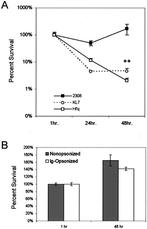
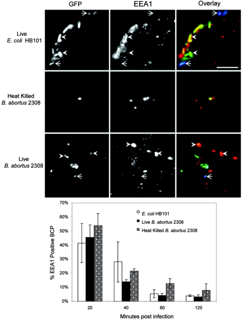
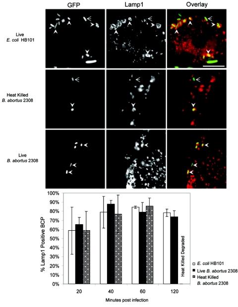
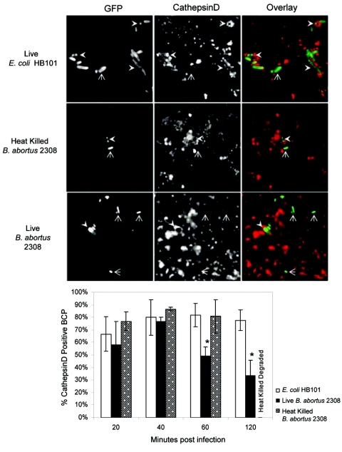
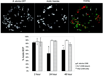
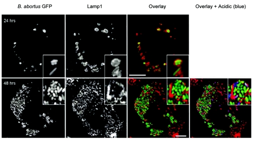
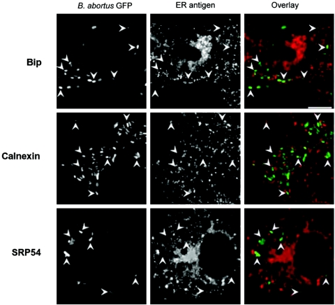
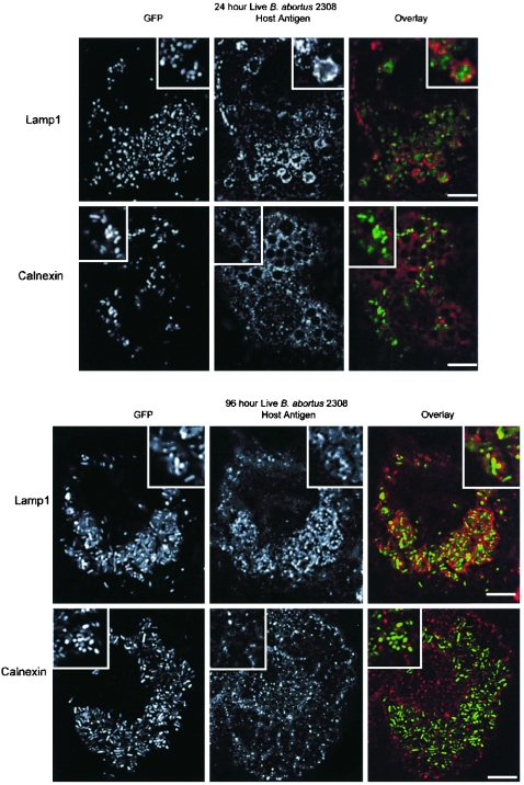
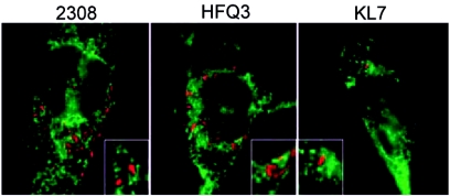
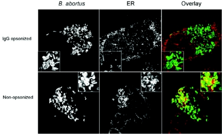
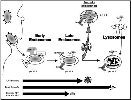
Cells in the Brucella spp. are intracellular pathogens that survive and replicate within host monocytes. Brucella maintains persistent infections in animals despite the production of high levels of anti-Brucella-specific antibodies. To determine the effect of antibody opsonization on the ability of Brucella to establish itself within monocytes, the intracellular trafficking of virulent Brucella abortus 2308 and attenuated hfq and bacA mutants was followed in the human monocytic cell line THP-1. Early trafficking events of B. abortus 2308-containing phagosomes (BCP) were indistinguishable from those seen for control particles (heat-killed B. abortus 2308, live Escherichia coli HB101, or latex beads). All phagosomes transiently communicated the early-endosomal compartment and rapidly matured into LAMP-1(+), cathepsin D(+), and acidic phagosomes. By 2 h postinfection, however, the number of cathepsin D(+) BCP was significantly lower for live B. abortus 2308-infected cells than for either Brucella mutant strains or control particles. B. abortus 2308 persisted within these cathepsin D(-), LAMP-1(+), and acidic vesicles; however, at the onset of intracellular replication, the numbers of acidic B. abortus 2308 BCP decreased while remaining cathepsin D(-) and LAMP-1(+). In contrast to B. abortus 2308, the isogenic hfq and bacA mutants remained in acidic, LAMP-1(+) phagosomes and failed to initiate intracellular replication. Notably, markers specific for the host endoplasmic reticulum were absent from the BCPs throughout the course of the infection. Thus, opsonized B. abortus in human monocytes survives within phagosomes that remain in the endosomal pathway and replication of virulent B. abortus 2308 within these vesicles corresponds with an increase in intraphagosomal pH.


Figures

FIG. 1.
FIG. 1.
Survival and replication of B. abortus 2308 (▪), KL7 (2308 bacA) (○), and Hfq3 (2308 hfq) (□) in THP-1 cells. (A) PMA-differentiated THP-1 cells were infected with IgG-opsonized B. abortus and incubated for the indicated times whereby monocytes were lysed and intracellular bacteria were enumerated by growth on BA plates following serial dilution. Results are presented as percent survival, which was calculated by dividing the number of intracellular brucellae present at the times indicated postinfection by the number of CFU at 1 h postinfection and multiplying by 100. The number of intracellular brucellae present at each time point represents the average of three wells of THP-1 cells infected with each individual strain. Results presented are of a single representative experiment chosen from three independent experiments. (B) Similar experiments were performed by infecting THP-1 cells with either Ig-opsonized or nonopsonized B. abortus 2308. Ig-opsonized bacteria were internalized at a level 10 times that observed for nonopsonized bacteria, and the difference in uptake was accounted for by calculating percent survival. Statistical significance was calculated using Student's t test analysis (**, P ≤ 0.001).
FIG. 2.
FIG. 2.
Transient association of Brucella-containing phagosomes (BCP) with the early-endosomal marker EEA1. Monocytes were infected with opsonized bacteria, fixed at times indicated, and scored for the percent localization of EEA1 antigen on BCP following immunofluorescence staining. Representative images of BCP from monocytes infected with either control particles (heat-killed B. abortus or E. coli HB101) or virulent live B. abortus cells are shown from the 20-min time point coinciding with the highest level of EEA1 colocalization. Identical magnification of images was used to illustrate the size difference between Brucella and E. coli (bar, 10 μ). Intracellular bacteria (solid arrow, only green fluorescence) were differentiated from extracellular bacteria (line arrow, green and blue fluorescence) by differential antibody staining (see Materials and Methods). Percent colocalization for EEA1 was performed by scoring BCP for the presence or absence of EEA1, and results shown represent the averages and standard deviations from three independent experiments. All three BCP transiently associated with EEA1 soon after internalization, followed by rapid loss of this marker. No statistical differences were detected among the different BCP. The kinetics for the acquisition and loss of endocytic markers Rab5 and transferrin receptor followed the same transient pattern described here for EEA1 (data not shown).
FIG. 3.
FIG. 3.
Rapid acquisition of phagosomal marker LAMP-1. Monocytes were infected with opsonized bacteria, and at times indicated in the graph, BCP were scored for the presence (indicated by solid arrow) or absence (indicated by line arrow) of LAMP-1. Representative images are shown at 60 min postinfection coinciding with peak LAMP-1 acquisition for all BCP types (bar, 10 μm). A significant reduction in detectable GFP for heat-killed BCP occurred at 2 h due to the fixation process (methanol) needed for optimal anti-LAMP-1 antibody staining. As a result, the numbers of detectable heat-killed Brucella cells visualized at 2 h were significantly lower than those for live virulent Brucella and E. coli and thus, colocalization percentages were not calculated for heat-killed bacteria at 2 h. Percent colocalization and standard deviations were calculated using results from three independent experiments. No statistical differences were detected among the different BCP types at any time point.
FIG. 4.
FIG. 4.
Early B. abortus-containing phagosomes are acidic. Monocytes were infected with bacteria for the times indicated in the graph and subsequently treated with the acidotropic dye Lysotracker Red prior to fixation. GFP-positive phagosomes were then scored on the basis of accumulation of the red acidotropic dye to calculate percent acidic BCP (positive, solid arrow; negative, line arrow). Visualization of acidotropic dye does not require methanol treatment that reduced the detection of dead Brucella GFP; therefore, adequate amounts of dead bacteria were able to be visualized at the 2-h time point, unlike samples analyzed for LAMP-1 or cathepsin D colocalization. Representative images are shown from samples collected at the 60-min time point. Colocalization percentages and standard deviations were calculated using results from three independent experiments. No statistical differences were detected among the different BCP types at any time point.
FIG. 5.
FIG. 5.
Early but not late B. abortus-containing phagosomes associate with lysosomes. Monocytes were incubated at the indicated times following infection with either E. coli, dead B. abortus, or live B. abortus bacteria and then fixed with 4% paraformaldehyde for 10 min and permeabilized with ice-cold methanol for 1 min. After washing with cold PBS, monolayers were incubated with mouse anti-cathepsin D in BSP, followed with secondary anti-mouse Cy3-conjugated antibodies (red). Representative images shown are from 60 min postinfection. BCP were scored for the presence (solid arrows) or absence (line arrows) of cathepsin D to calculate percent colocalization. Results from three independent experiments were used to calculate the average and standard deviation for each time point. Statistical comparisons of live B. abortus to E. coli (60 and 120 min postinfection) and dead B. abortus (60 min postinfection) were significant by two-tailed Student's t test analysis (*, P ≤ 0.05).
FIG. 6.
FIG. 6.
B. abortus 2308 resides predominately in nonacidic compartments at 24 and 48 h postinfection in THP-1 cells, while isogenic bacA and hfq mutants remain largely confined to acidified phagosomes within these phagocytes. Only phagosomes containing single bacteria (BCP) were scored for the presence of the acidic marker (see text). Illustrated are panels from a representative macrophage scored for acidic (solid arrows) and nonacidic (line arrows) B. abortus 2308-containing phagosomes at 24 h postinfection. The results presented are averages from multiple independent experiments for each stain. *, P ≤ 0.05 for comparisons of phagosomes containing B. abortus 2308 with those containing B. abortus KL7 or Hfq3 using the Student's t test.
FIG. 7.
FIG. 7.
Replication of B. abortus 2308 within LAMP-1-positive vesicles that are largely in THP-1 cells at 48 h postinfection. Representative microscopic images of the infected THP-1 monolayers are shown during the replicative phase of the intracellular life cycle of B. abortus 2308 in these phagocytes. Acidified vesicles (blue) were labeled prior to fixation by treating cells with Lysotracker (Molecular Probes). Images were collected using a Bio-Rad 2000 scanning laser microscope. LAMP-1 primary antibodies were visualized using Cy5-conjugated anti-mouse secondary antibody (red fluorescence). Bar, 10 μm.
FIG. 8.
FIG. 8.
Proximal staining of ER markers and Brucella-containing phagosomes. Monocytes were fixed 2 h postinfection and stained for ER-specific antigens Bip, SRP54, and calnexin (red). Although few Brucella vesicles were seen positively colocalized with the host ER, the majority of Brucella-containing phagosomes were observed adjacent to areas of intense ER staining that was characterized as “proximal ER staining” and not positive colocalization of Brucella within the host ER network. Similar staining patterns, for both positive and proximal ER staining, were observed for latex beads, heat-killed Brucella, and avirulent E. coli (data not shown). Arrows note the position of Brucella-containing phagosomes to aid in comparing their location to ER markers. Bar, 5 μm.
FIG. 9.
FIG. 9.
Comparison of LAMP-1 and ER colocalization with Brucella-containing phagosomes in THP-1 monocytes at 24 and 96 h postinfection. Antibody-opsonized B. abortus 2308 cells (GFP) were used to infect monolayers for 24 and 96 h, fixed, and then stained for either LAMP-1 or calnexin host antigen (red). Phagosomes containing either single or multiple Brucella cells colocalized extensively with LAMP-1 at both 24 and 96 h and were identical to those obtained earlier with monolayers infected at 48 and 72 h postinfection with live B. abortus (Fig. 7). The tight and continuous opposition of LAMP-1 to intracellular Brucella is in contrast to the punctuated and more proximal staining pattern observed for the ER marker calnexin. A typical cluster from each micrograph was magnified to further illustrate the differences in staining patterns between LAMP-1 and calnexin markers (panel inserts). Staining for other ER-specific antigens (Bip, Sec61β, and SRP54) revealed similar patterns of ER proximal to Brucella phagosomes as well as latex beads (not shown). Bar, 10 μm.
FIG. 10.
FIG. 10.
Association of late-infection Brucella-containing phagosomes with the endoplasmic reticulum of THP-1 cells. THP-1 cells infected with B. abortus 2308, Hfq3 (2308 hfq), or KL7 (2308 bacA) were fixed at 48 h postinfection and stained for host ER elements using anti-Bip-specific antibody. Grayscale images were pseudocolored to make the GFP-labeled brucellae appear red, and Cy3-labeled Bip appears green. Consistent with the results obtained from brucellacidal assays employing these phagocytes (Fig. 1), considerably more brucellae were seen in THP-1 cell monolayers infected with virulent B. abortus 2308 at 48 h postinfection than were seen in THP-1 cells infected with B. abortus Hfq3 or KL7. A monocyte with a moderate bacterial burden of B. abortus 2308 was shown for a more accurate comparison to ER staining patterns observed for the attenuated mutants. No differences were detected, however, between these interactions of phagosomes containing these strains with the ER of THP-1 cells. Clear colocalization of ER markers with Brucella-containing vesicles was visible for some vesicles (filled arrows); however, the majority of the Brucella-containing phagosomes appear to be in close proximity to, but not contiguous with, the ER of THP-1 cells (line arrows) (see text). Arrows note the positions of Brucella-containing phagosomes to aid in comparing their location to ER markers. Bar within large panel, 10 μm; bar within panel insert, 1 μm.
FIG. 11.
FIG. 11.
Localization of nonopsonized B. abortus 2308 with the endoplasmic reticulum. THP-1-differentiated monocytes were infected with either Ig-opsonized or nonopsonized B. abortus. At 76 h postinfection, the monolayers were fixed and stained for the endoplasmic reticulum using anti-calnexin antibodies followed with Cy3-conjugated secondary antibodies (red). In the representative images shown, the Ig-opsonized bacteria were primarily found in vesicles that did not colocalize with the ER marker. In contrast, monocytes infected with nonopsonized B. abortus did contain an appreciable number of Brucella cells colocalizing with the ER marker. Although not all of the vesicles containing nonopsonized bacteria were positive for the ER, the presence of any distinct ER colocalization was absent from monocytes infected with opsonized bacteria.
FIG. 12.
FIG. 12.
Schematic representation of the intracellular trafficking patterns of IgG-opsonized B. abortus 2308, KL7 (2308 bacA), and Hfq3 (2308 hfq) in the human monocytic cell line THP-1. The black arrows at the bottom of the figure denote the extent to which phagosomes containing the different B. abortus strains progress down the endolysosomal pathway within these host cells. All Brucella cells were trafficked rapidly through the early- and late-endosomal compartments regardless of viability or virulence status. Progression of phagosomes beyond the late-endosomal compartment (LAMP-1+ and acidic) correlated with the viability/virulence status of the bacteria used for infection (see Discussion). Remaining viable Brucella cells were found within phagosomes that were no longer acidic (pH ≥ 6) while remaining LAMP-1 positive and cathepsin D negative. Replicating Brucella cells were also observed within this same nonacidic compartment, demonstrating a correlation between the rise in intraphagosome replication and the shift in bacterial physiology from survival to intracellular replication (speckled arrow). By contrast, phagosomes containing attenuated Brucella Hfq3 and KL7 were unable to progress to either fuse with lysosomes or take on the characteristics of a replicative vesicle while phagosomes containing heat-killed Brucella proceeded to fuse with lysosomes unabated at the same rates as phagosomes containing avirulent E. coli and latex beads.

Similar articles

Cited by

References

    1. Arenas, G. N., A. S. Staskevich, A. Aballay, and L. S. Mayorga. 2000. Intracellular trafficking of Brucella abortus in J774 macrophages. Infect. Immun. 68:4255-4263. - PMC - PubMed
    1. Baldwin, C. L., and M. Parent. 2002. Fundamentals of host immune response against Brucella abortus: what the mouse model has revealed about control of infection. Vet. Microbiol. 90:367-382. - PubMed
    1. Baldwin, C. L., and A. J. Winter. 1994. Macrophages and Brucella. Immunol. Ser. 60:363-380. - PubMed
    1. Boschiroli, M. L., S. Ouahrani-Bettache, V. Foulongne, S. Michaux-Charachon, G. Bourg, A. Allardet-Servent, C. Cazevieille, J. P. Liautard, M. Ramuz, and D. O'Callaghan. 2002. The Brucella suis virB operon is induced intracellularly in macrophages. Proc. Natl. Acad. Sci. USA 99:1544-1549. - PMC - PubMed
    1. Celli, J., C. de Chastellier, D. M. Franchini, J. Pizarro-Cerda, E. Moreno, and J. P. Gorvel. 2003. Brucella evades macrophage killing via VirB-dependent sustained interactions with the endoplasmic reticulum. J. Exp. Med. 198:545-556. - PMC - PubMed

Publication types

LinkOut - more resources