. 2005 Jul 22;280(29):27085-92.
doi: 10.1074/jbc.M409567200. Epub 2005 May 20.

Keratocan expression of murine keratocytes is maintained on amniotic membrane by down-regulating transforming growth factor-beta signaling

Affiliations

Keratocan expression of murine keratocytes is maintained on amniotic membrane by down-regulating transforming growth factor-beta signaling

Tetsuya Kawakita et al. J Biol Chem. .

Abstract

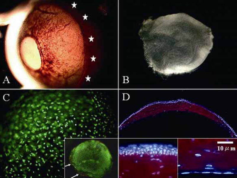
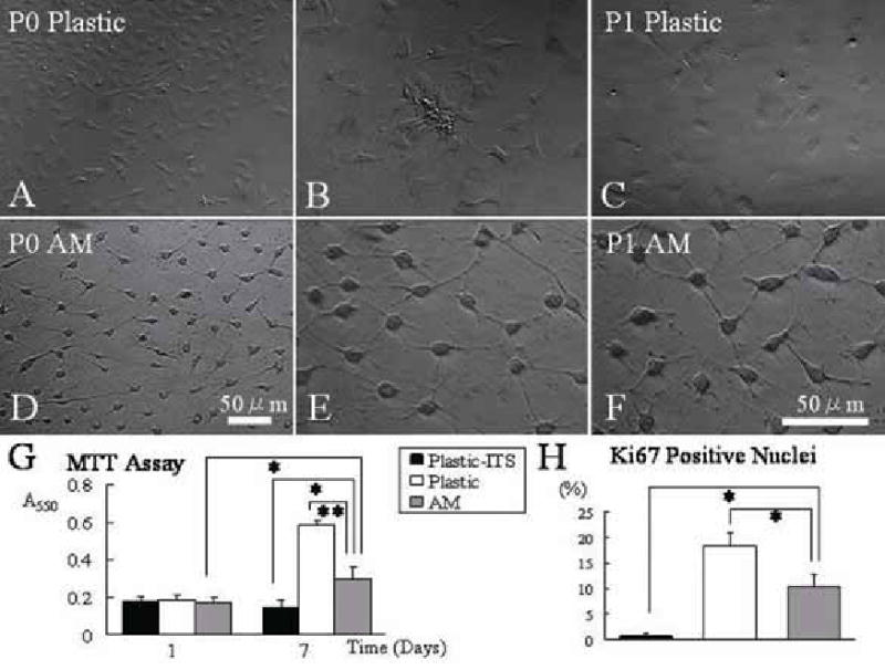
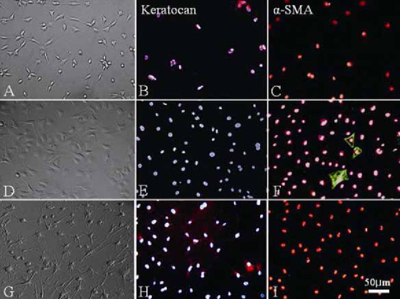
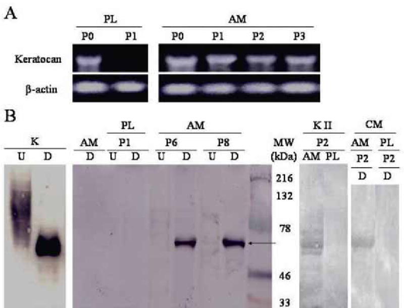
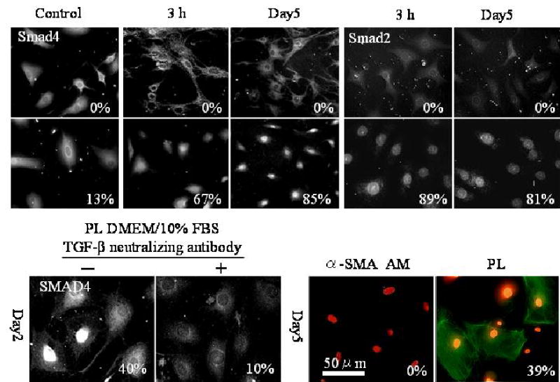
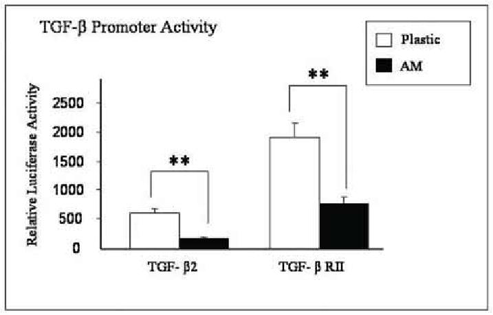
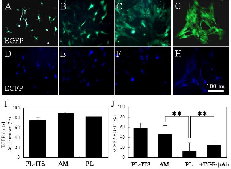
Keratocytes in the corneal stroma express keratan sulfate-containing proteoglycans including cornea-specific keratocan. On plastic dishes, human, bovine, and rabbit keratocytes lose their characteristic dendritic morphology and keratocan expression when cultured in serum-containing media. Herein, we demonstrated that murine keratocytes also acquired a fibroblastic shape and lost keratocan expression after first passage when cultured on plastic in the presence of serum. In contrast, cells expanded on human amniotic membrane (AM) stromal matrix maintained a three-dimensional dendritic morphology and expressed keratocan mRNA and protein for at least 8 passages before senescence. When keratocytes were cultured on AM, the promoter activity of transforming growth factor (TGF)-beta2 and TGF-beta receptor II was down-regulated as compared with that on plastic. Furthermore, cells on AM continuously retained Smad 2 and Smad 4 in the cytoplasm and did not express alpha-smooth muscle actin, even when 10 ng/ml TGF-beta1 was added in a serum-free medium for up to 5 days. In parallel to such down-regulation of TGF-beta signaling, keratocan promoter-driven ECFP expression was observed in cells cultured either on AM in the presence of serum or on plastic containing serum treated with a neutralizing antibody to TGF-beta. Collectively, these results indicate that down-regulation of Smad-mediated TGF-beta signaling is an important mechanism for cultured keratocytes to maintain a normal phenotype while continuously expanded in a serum-containing medium. This strategy of suppressing TGF-beta signaling, achieved by AM stromal matrix in part via suppression of TGF-beta gene transcription, can be used to expand keratocytes in culture without the use of AM in the future.


Figures

Fig. 1
Fig. 1
In vivo keratocyte morphology and keratocan expression. An enucleated CD-1 albino mouse eye shows a distinguishable limbal region under transillumination (A, marked by stars). The corneal stroma is dissected from the adjacent sclera (see Methods) (B). Under high magnification, Live/Dead assay® shows a dendritic morphology of all viable (stained green) keratocytes distributed in a three dimension manner (more visible on the plane in focus) (C). Dead cells (stained red) are found only in the excised stromal edge (C, inset, white arrows). Immunostaining demonstrates keratocan expression exclusively in the corneal stroma (D). Corneal epithelial cells and endothelial cells do not express keratocan (D, inset).
Fig. 2
Fig. 2
Cellular morphology and proliferation on plastic and AM cultures. Passage 0 (P0) cells expanded on plastic are spindle-shaped with a broad satellite cytoplasm (A and B), and become more flattened after subcultured to P1 (C). In contrast, cells on AM maintain a triangular shaped cell body and a scanty cytoplasm with dendritic processes (D and E) even after subcultured to P1 (F). Extensive intercellular cell-cell contacts are evident in cells expanded on AM. Micrograph A and D are taken at same magnification and B to F at higher magnification. Both MTT assay (G) and quantitation of Ki67 nuclear staining (H) show that cells on AM in DMEM/10% FBS had a higher proliferative activity than cells on plastic in DMEM/ITS, but less than cells on plastic in DMEM/10% FBS (*p<0.05 and **p<0.01, for both G and H).
Fig. 3
Fig. 3
Keratocan and α-SMA expression. On plastic, cells of primary cultures (P0) in serum-free DMEM/ITS have a dendritic morphology (A) and expressed keratocan (B), but not α-SMA (C). Cells cultured in DMEM/10% FBS expand in number (D) but completely lose keratocan expression (E), while some cells express α-SMA (F). In contrast, P2 cells expanded on AM in DMEM/10% FBS continue to be dendritic, form cell-cell networks (G) similar to P0 cultures, and express keratocan (H), but not α-SMA (I).
Fig. 4
Fig. 4
CD34 and fibronectin expression. CD34 is not expressed (A), but fibronectin is expressed intracellularly and extracellularly (B) by P2 cells cultured on plastic. In contrast, CD34 is maintained (C) while fibronectin is not expressed (D) by P2 cells cultured on AM.
Fig. 5
Fig. 5
Expression of keratocan transcript and proteins. (A) Expression of keratocan transcript (size of 1065bp) is maintained in AM cultures from passage 0 (P0) to passage 3 (P3). In contrast, such expression is maintained on plastic at P0, but lost at P1. β -actin was used as a loading control. (B) Cornea stroma extracted with 4 M guanidine-HCl from the normal corneal stroma (K), cells cultured on AM at P6 and P8 (designated as A6 and A8, respectively), and cell cultured on plastic at P1 (designated as P1) are treated with (D) or without (U) endo-β-galactosidase to remove keratan sulfate, and subjected to Western blot using an antibody to murine keratocan. The undigested samples of the normal cornea and A6 and A8 show high MW smearing, while the digested counterparts reveal a 50 kD band of keratocan (arrow). A similar 50 kDa band was obtained from P2 cultures on AM, but not from P2 cultures on plastic by keratanase II digestion (KII, P2, AM and PL). AM alone without cells (AM) does not express keratocan. The digested sample of the conditioned media of P2 AM cultures, but not those of P2 plastic cultures, also shows a 50 kD band.
Fig. 6
Fig. 6
Suppression of Smad-mediated TGF-β signaling by AM. (A) Immunostaining shows that Smad 4 nuclear translocation takes place only in a small percentage of cells (13%) cultured on plastic in DMEM/ITS, but increased to 67% and 85% after 10 ng/ml TGF-β1 is added for 3 h. and 5 days, respectively. A similar trend is noted for Smad 2 nuclear localization. (B) Nuclear localization of Smad 4 is noted in 40% of cells on plastic in DMEM/10% FBS, but in 10% of cells when TGF-β neutralizing antibody is added for two days. (C) Expression of α-SMA is noted in 39% of cells cultured on plastic after 10 ng/ml TGF-β1 for 5 days, but is negative in cells cultured on AM.
Fig. 7
Fig. 7
Suppression of TGF-β promoter activity by AM. Mouse corneal fibroblasts (P1) cultured on plastic and AM are cotransfected with TGF-β2 promoter-luc plus CMV-β-galactosidase or TGF-βRII promoter–luc plus CMV-β-galactosidase for 48 hours. Cell extracts are assayed for both activities of luciferase and β-galactosidase. The relative luciferase unit of the promoter activity of TGF-β2 and TGF-βRII is suppressed in cells cultured on AM (** p < 0.01).
Fig. 8
Fig. 8
Keratocan promoter activity. Aden-track-Kerapr3.2-intron-ECFP/BpA adenovirus (50 M.O.I.) is used to transfect primary cultures in DMEM/ITS on plastic (A), DMEM/10% FBS on AM (B), and DMEM/10% FBS on plastic without (C) or with TGF-β neutralizing antibody (D). Transfection efficiency determined by EGFP expression (green fluorescence) is more than 80 % in these experiements (I). On the same field, cells expressing EGFP was then analyzed for keratocan promoter activity by monitoring ECFP expression (blue fluorescence). Keratocan expression is noted in 59 ± 9.5 % cells cultured on plastic in DMEM/ITS (E) and 46 ± 6.2 % of cells on AM cultures in DMEM/10% FBS (F), and is significantly reduced in cells cultured on plastic in DMEM/10% FBS (13 ± 2.6%, G), (**p<0.01, J), but is partially restored to 25 ± 5.6 % (**p<0.01, J) when added with a TGF-β neutralizing antibody (H).

Similar articles

Cited by

References

    1. Beals MP, Funderburgh JL, Jester JV, Hassell JR. Invest Ophthalmol Vis Sci. 1999;40:1658–1663. - PubMed
    1. Jester JV, Barry-Lane PA, Cavanagh HD, Petroll WM. Cornea. 1996;15:505–516. - PubMed
    1. Espana EM, He H, Kawakita T, Di Pascuale MA, Raju VK, Liu CY, Tseng SC. Invest Ophthalmol Vis Sci. 2003;44:5136–5141. - PubMed
    1. Espana EM, Kawakita T, Liu CY, Tseng SCG. Invest Ophthalmol Vis Sci. 2004;45:2985–2991. - PubMed
    1. Dahl IM, Johnsen W, Anseth A, Prydz H. Exp Cell Res. 1974;88:193–197. - PubMed

Publication types

MeSH terms

LinkOut - more resources