. 2005 Sep;289(3):H1099-105.
doi: 10.1152/ajpheart.00423.2005. Epub 2005 May 20.

Electromechanical analysis of infarct border zone in chronic myocardial infarction

Affiliations

Electromechanical analysis of infarct border zone in chronic myocardial infarction

Hiroshi Ashikaga et al. Am J Physiol Heart Circ Physiol. 2005 Sep.

Abstract

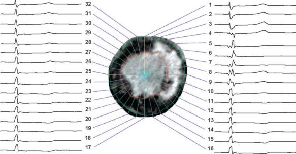
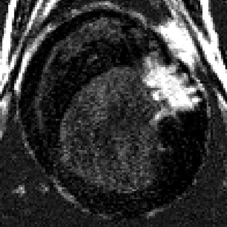
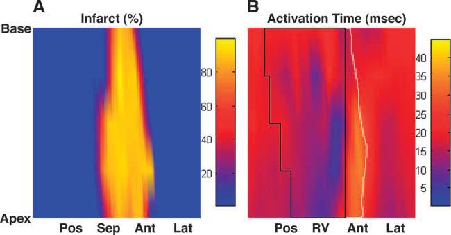
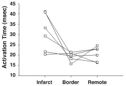
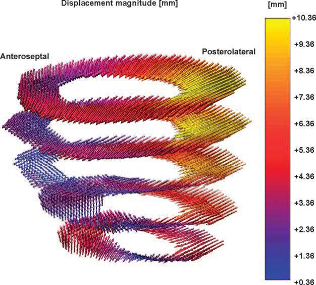
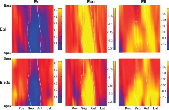
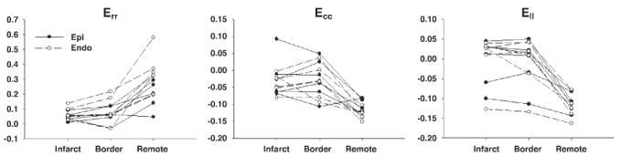
To test the hypothesis that alterations in electrical activation sequence contribute to depressed systolic function in the infarct border zone, we examined the anatomic correlation of abnormal electromechanics and infarct geometry in the canine post-myocardial infarction (MI) heart, using a high-resolution MR-based cardiac electromechanical mapping technique. Three to eight weeks after an MI was created in six dogs, a 247-electrode epicardial sock was placed over the ventricular epicardium under thoracotomy. MI location and geometry were evaluated with delayed hyperenhancement MRI. Three-dimensional systolic strains in epicardial and endocardial layers were measured in five short-axis slices with motion-tracking MRI (displacement encoding with stimulated echoes). Epicardial electrical activation was determined from sock recordings immediately before and after the MR scans. The electrodes and MR images were spatially registered to create a total of 160 nodes per heart that contained mechanical, transmural infarct extent, and electrical data. The average depth of the infarct was 55% (SD 11), and the infarct covered 28% (SD 6) of the left ventricular mass. Significantly delayed activation (>mean + 2SD) was observed within the infarct zone. The strain map showed abnormal mechanics, including abnormal stretch and loss of the transmural gradient of radial, circumferential, and longitudinal strains, in the region extending far beyond the infarct zone. We conclude that the border zone is characterized by abnormal mechanics directly coupled with normal electrical depolarization. This indicates that impaired function in the border zone is not contributed by electrical factors but results from mechanical interaction between ischemic and normal myocardium.


Figures

Fig. 1
Fig. 1
Epicardial electrical signals during a cardiac cycle (cycle length = 544 ms, heart rate = 110 beats/min) registered to a short-axis delayed hyperenhancement (DHE) MR image of the left ventricle (LV) split into 32 sectors. Regions of bright intensity correspond to myocardial infarction (MI). In this image, abnormal or delayed activation is seen in sectors 4 through 9, whereas the infarct ranges from sector 3 through sector 14.
Fig. 2
Fig. 2
Short-axis DHE image of MI from a single animal. Regions of bright intensity correspond to MI. Note multiple interdigitations of viable myocardium within the infarct region.
Fig. 3
Fig. 3
Infarct depth and electrical activation from a single animal. A: infarct map (in %depth). B: isochrone map of electrical activation time (in ms). The area circumscribed by a solid black line represents the right ventricle (RV). The area circumscribed by a solid white line represents the infarct zone in the anterior wall (the infarct zone in the septum is covered by the RV). Pos, posterior wall; Sep, septal wall; Ant, anterior wall; Lat, lateral wall.
Fig. 4
Fig. 4
Quantitative analysis of electrical activation time in each zone (n = 6). Electrical activation time (in ms) was significantly increased in the infarct zone compared with that in the border zone [31 (SD 9) vs. 19 (SD 2) ms, P < 0.05], whereas it was not significantly different between the border zone and the remote zone [19 (SD 2) vs. 21 (SD 3) ms, P = not significant (NS)].
Fig. 5
Fig. 5
Three-dimensional (3D) displacement map from a single animal. Each arrow represents a displacement vector that points from the end-diastolic to the end-systolic configuration. The magnitude of displacement is color coded. It is clear that the displacement magnitude in the infarct zone in the anteroseptal wall (left) is small (purple to blue) compared with that of the remote zone in the posterolateral wall (right, red to yellow).
Fig. 6
Fig. 6
3D strain map from a single animal. The area circumscribed by a solid white line represents the infarct zone. Radial (Err), circumferential (Ecc), and longitudinal (Ell) strains are shown. The regions of abnormal strains, particularly Ell, extended far beyond the infarct zone, and the strains were larger in the epicardium than in the endocardium. Epi, epicardial layer; Endo, endocardial layer.
Fig. 7
Fig. 7
Quantitative analysis of finite strain in each zone (n = 6). Err, Ecc, and Ell in both the infarct zone and the border zone were significantly smaller than those in the remote zone (P < 0.05), and there was no significant difference between the infarct zone and the border zone (P = NS). There was a significant transmural gradient between the epicardium and endocardium in Err, Ecc, and Ell in the remote zone (P < 0.05), and the transmural gradient was lost in both the infarct zone and the border zone.

References

    1. Aletras AH, Ding S, Balaban RS, Wen H. DENSE: displacement encoding with stimulated echoes in cardiac functional MRI. J Magn Reson. 1999;137:247–252. - PMC - PubMed
    1. Aletras AH, Wen H. Mixed echo train acquisition displacement encoding with stimulated echoes: an optimized DENSE method for in vivo functional imaging of the human heart. Magn Reson Med. 2001;46:523–534. - PMC - PubMed
    1. Barr RC, Spach MS. Sampling rates required for digital recording of intracellular and extracellular cardiac potentials. Circulation. 1977;55:40–48. - PubMed
    1. Cohn JN. Post-MI remodeling. Clin Cardiol. 1993;16:II21–II24. - PubMed
    1. Curran-Everett D, Benos D. Guidelines for reporting statistics in journals published by the American Physiological Society. Am J Physiol Heart Circ Physiol. 2004;287:H447–H449. - PubMed

Publication types